Download
1 / 41
450 likes | 1.05k Views
PATHOLOGY OF ADRENAL GLANDS. Department of Internal Medicine №2 as.-prof. Martynyuk L.P. Plan of lecture. Anatomy of adrenal glands Regulation of hormone’s secretion Biological effects of adrenal gland hormones Chronic adrenal failure: diagnostic criteria, treatment. Adrenal crisis

E N D
PATHOLOGY OF ADRENAL GLANDS Department of Internal Medicine №2 as.-prof. Martynyuk L.P.
Plan of lecture • Anatomy of adrenal glands • Regulation of hormone’s secretion • Biological effects of adrenal gland hormones • Chronic adrenal failure: diagnostic criteria, treatment. • Adrenal crisis • Pheochromocynoma : diagnostic criteria, treatment • Cushing’s syndrome : diagnostic criteria, treatment • Hypothalamic syndrome of puberty period : diagnostic criteria, treatment
Historical perspectives • 1563: Eustachius first described adrenal glands • 1855: Thomas Addison noted real importance of adrenal glands • 1856: Brown Sequard demonstrated that adrenals are very necessary for life (without adrenal glands animals could not survive)
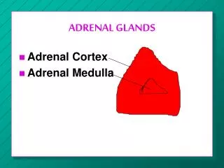
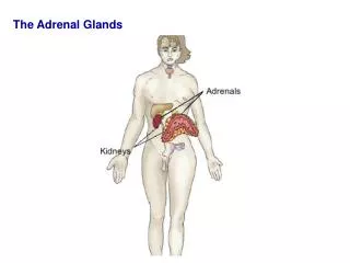
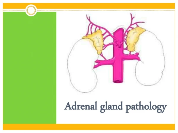
Anatomy of adrenal glands Localization: the top of the kidney and weighting approximately 5 g each. Vascularization: a. suprarenalis superior (from a. phrenica inferior), a. suprarenalis media (from aorta abdominalis), a suprarenalis inferior (from a. renalis). Innervation: n. splanchnicus major (through plexus celiacus and plexus renalis), fibrae n. vagus and n. phrenicus.
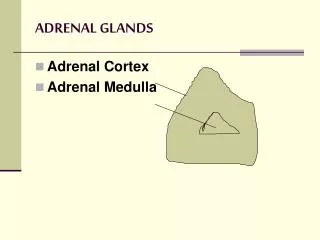
Adrenal cortex include three zones: • Glomerular (glomerulosa) produces mineralocorticoids (e.g., aldosterone); • Fascicular (fasciculata) produces glucocorticoids (e.g., cortisol (hydrocortisone)); • Reticular (reticularis) produces cortisol and androgens (dehydroisoandrosterone (dehydroepiandrosterone)), which exert their chief physiologic activity after conversion to testosterone and dehydrotestosterone. Adrenal medulla • produces catecholamines.
Action of mineralocorticoids: regulation of electrolyte balance in the organism: • increasing the level of sodium (by sodium retention in distal nephron, colon, salivary gland) • decreasing the level of potassium (by excretion).
Action of glucocorticoids: • increasing of glycogen synthesis in liver and decreasing of glucose utilization by peripheral tissues, increasing gluconeogenesis; • increasing of protein synthesis in liver and decreasing of its synthesis in muscles and increasing of protein destruction in muscles; • increasing of lipolisis; • anti-inflammatory function and immunomodulation; • cardiovascular regulation (increasing of blood pressure).
Regulation of secretion • glucocorticoids’ and androgens’ secretion is regulated by hypothalamic – pituitary system • mineralocorticoids’ secretion is regulated by the renin – angiotensin system, the level of Na+, K+ in blood, and to a lesser extent of ACTH
Catecholamines are produced from the tyrosine(organism takes it from the meal or from the phenilalanine in the liver) → dioxyphenilalanine (DOPHA) → dopamine (it goes into blood only from some neurons of the central nervous system) → norepinephrine(noradrenaline) (it goes into blood only from sympathetic teleneurons) → epinephrine(adrenaline) (it goes into blood only from adrenal medulla). The principle urinary metabolic products of epinephrine and norepinephrine are the metanephrines and vanillylmandalic acid (VMA).
CHRONIC ADRENOCORTICAL INSUFFICIENCY. It is an insidious and usually progressive disease resulting from adrenocortical hypofunction.
Classification. • Primary adrenocortical insufficiency (Addison’s disease). • Secondary adrenocortical insufficiency .
Etiology of adrenal insufficiency: Primary: • autoimmune processes (50 – 65 %); • tuberculosis; • neoplasm, metastatic carcinoma; • inflammatory necrosis; • amyloidosis; heamochromatosis; • bilateral adrenal hemorrhage or infarction, intra – adrenal hemorrhage (Waterhouse – Friedrichsen syndrome following meningococcal septicemia); • bilateral adrenalectomy; Secondary: • hypothalamic or pituitary disease (primary injury of these organs leads to insufficiency of ACTH secretion that cause the two – side atrophy of adrenal glands); • glucocorticoid therapy.
Pathogenesis. Deficiency of adrenal hormones contributes to the hypotension and produces disturbances in carbohydrate, fat, and protein metabolism, and severe insulin sensitivity.
Symptoms and signs. Presentation may be acute and chronic. Frequently clinical signs of the primary chronic adrenocortical insufficiency are manifested in that time when adrenocortical tissue is destroyed on 70-90 %. The most common complaints are: • weakness, • malaise, • weight loss, • anorexia, • Depression, • Hyperpigmentation of the skin
Objective examination: • Hypotension or postural hypotension • Tachycardia. • Weight loss • Anorexia, nausea, vomiting, abdominal pain, diarrhea are often. Gastritis, ulcer disease can occur. • Decreased cold tolerance, with hypometabolism may be noted. • Sexual disorders. • Neurologic and psychiatric disorders: • Hypoglycemia.
Laboratory findings. • A low serum Na level and a high serum P level • Adrenal insufficiency can be specifically diagnosed by: • low levels of plasma glucocorticoids and mineralocorticoids, or urinary 17 – hydroxycorticosteroid (17 – OHCS) or 17 – ketogenic steroid (17 – KGS); • demonstrating failure to increase plasma cortisol levels, or urinary 17 – OHCS or 17 – KGS excretion, upon administration of ACTH • To distinguish between primary and secondary adrenal insufficiency, me have to find the level ofplasma ACTH: primary shows increased, and secondary shows decreased level.
Instrumental findings • The ECG may decreased voltage and prolonged P – R and Q – T intervals. • The EEG shows alized slowing of the α – rhythm.
Treatment. • Etiologic: appropriate treatment of complicating infections (e.g., tuberculosis). • Pathogenic: • Diet (enough quantity of proteins, vitamins, salt and water). • Glucocorticoids Average dosage is: • cortisol: 20 – 25 mg daily; • prednisolone 5 – 7.5 mg daily; • hydrocortisone 30 – 40 mg orally daily.
Mineraloocorticoids. DOCSA 5 mgorally daily should be used in patients with severe and moderate duration orfludrocortisone 0.1 – 0.2 mgorally once a day is recommended • Intercurrent illnesses (e.g., infections) should be regarded as potentially serious and the patient should double his dosage until he is well. • If nausea or vomiting preclude oral therapy, medical attention should be sought immediately and parental therapy started.
Adrenal crisis - is a medical emergency caused by sudden marked insufficiency of adrenocortical hormones.
Precipitating factors • stress (infection (especially with septicemia), trauma, surgery, prolonged fasting, salt loss due to excessive sweating during hot weather); • sudden withdrawal of adrenocortical hormone therapy in patients with chronic insufficiency.
Clinical features. An adrenal crisis is characterized by • profound asthenia, • severe pains in the abdomen, lower back or legs; • nausea, vomiting diarrhea; • peripheral vascular collapse; • renal shutdown with azotemia. • Body temperature may be subnormal, through severe hyperthermia due to infection is often seen.
Treatment. Therapy should be instituted immediately once a provisional diagnosis of adrenocortical failure has been made. • Substitution therapy • Rehydration • Treatment of complications (hyperpyrexia, psychotic reactions).
PHEOCHROMOCYTOMA. It is a tumor of chromaffin cells that secrete catecholamines
Clinical features • hypertension, • Tachycardia, diaphoresis, postural hypotension, tachypnea, • flushing, • cold and clammy skin, severe headache, angina, palpitation, • visual disturbances, dyspnea, parasthesias • nausea, vomiting, epigastric pain, constipation or diarrhea and a sense of impending doom are common; some or all of these symptoms and signs may occur in any patient.
Investigations • An increased 3-h (24-h) urinary excretion of epinephrine, norepinephrine and their metabolic products (VMA or metanephrines). • Increased plasma epinephrine, norepinephrine. • CT scanning of the abdomen for the localization of the tumor. • Scanning is useful for extra – adrenal tumors.
Treatment • Surgical removal of the tumor is the treatment of choice. • During crisis a combination of α- and β- adrenergic blocking agents • phentolamine (tropaphen) 2 - 4 mg every 5 - 10 min till stopping of the crisis, • phenoxybenzamine 10 – 20 mg 3 – 4 times daily, • propranolol 30 – 60 mg/day • and infusion of sodium nitroprusside.
Cushing's syndrome is a constellation of signs and symptoms caused by prolonged excessive amounts of circulating cortisol.
Clinical features • Obesity Typically the distribution of fat involves the trunk, particularly the cervicodorsal region (buffalo hump), supraclavicular area, and abdomen. The face is round and plethoric (moon face). The extremities are thin in relation to the rest of the body.
The skin of patients with Cushing's syndrome is thin and fragile, ecchymoses and hematomas results from a combination of thin skin and capillary fragility. • Striae, when present, are usually located on the abdomen, breast, and axillae. Occasionally, they may be found on the back and on the extremities. They are pink or purplish in color and wider than 1 cm
Clinical features • Hypertension • Proximal muscular weakness affecting predominantly the muscles of the pelvic girdle • Acne, hirsutism, and menstrual irregularities • Osteoporosis, compression fractures and persistent back pain • Peripheral edema • psychiatric manifestations, including anxiety, depression, and even frank psychosis
Diagnosis. There are two phases of investigation: • confirmation of the presence or absence of Cushing’s syndrome; • differential diagnosis of its case.
Glucose intolerance, glucosuria is often seen, but ketoacidosis or the chronic complications of hyperglycemia are very uncommon • The level of cortisol and ACTH • Dexamethasone suppression test • Radiologic diagnosis includes X-ray examination for a pituitary tumor, and computed tomography which is the most popular procedure for visualizing the adrenals in patients with Cushing's syndrome
Treatment. 1) Surgery: • the transfrontal exploration, • transphenoidal hypophysectomia • adrenalectomy 2) pituitary irradiation. 3) pharmacologic therapy • peritol (4 mg 2 - 3 times a day, which is increased to a maximum dose of 4 mg every 4 h over 2 to 4 – 6 – 12 weeks • the dopamine agonist bromocriptine the usual dose is 2.5 mg three times a day. But the therapy has to be began from the ¼ of a tablet (2.5 mg) at a bedtime for 3 to 4 days (because of its side effect such as somnolence), then it has to be increased on ¼ of a tablet each 3 days to 7.5 mg. • symptomatic therapy
Hypothalamic syndrome of pubertal period. Particularities. • Obesity is not cushingoid (not central). • Striae (pink and not very large). • Hypertension (constant or permanent). • Glucose intolerance.
Treatment. • Diet 8. • Parlodel (2.5 – 5 mg for 3 – 6 month). • Peritol (4 mg 2 times a day for 1 month). • Dehydration therapy (hypothiasid 50 – 100 mg/day MgSO4 25 % solution intramuscular 10 – 15 times). • Nonsteroid anti-inflammatory drugs (indometacine). • Biogenic stimulators (aloe, plasmol). • Increasing of microcirculation of the blood in the brain (cavinton, piracetam). • Vitamintherapy. • Symptomatic therapy (hypotensive therapy). • Physiotherapy.
References • The Merck Manual of Diagnosis and Therapy (fourteenth Edition)/ Robert Berkow and others. – published by Merck Sharp & Dohme Research Laboratories, 1982. – P. 1014 – 1019,1025 – 1028, 1021 – 1024. • Manual of Endocrinology and Metabolism (Second Edition)/ Norman Lavin. – Little, Brown and Company.- Boston-New York-Toronto-London, 1994. - P. 111 – 142, 173 - 180.. • Endocrinology (A Logical Approach for Clinicians (Second Edition)). William Jubiz.-New York: WC Graw-Hill Book, 1985. - P. 38 – 42, 144 –164, 198 – 205. • Short Textbook of Medical Diagnosis and Management (Third Edition). Mohammad Inam Danish. – Pakistan, 2002. – P.459 – 462, 504 – 505.