Download
1 / 72
890 likes | 1.45k Views
Diseases of adrenal glands and hypophysis. Е thiology, pathogenesis, Diagnostics. Clinical pattern. Complications. Principles of treatment. The role of a doctor-dentist in early diagnostics and prophylaxis. Anatomy of adrenal glands.

E N D
Diseases of adrenal glands and hypophysis. Еthiology, pathogenesis, Diagnostics. Clinical pattern. Complications. Principles of treatment. The role of a doctor-dentist in early diagnostics and prophylaxis.
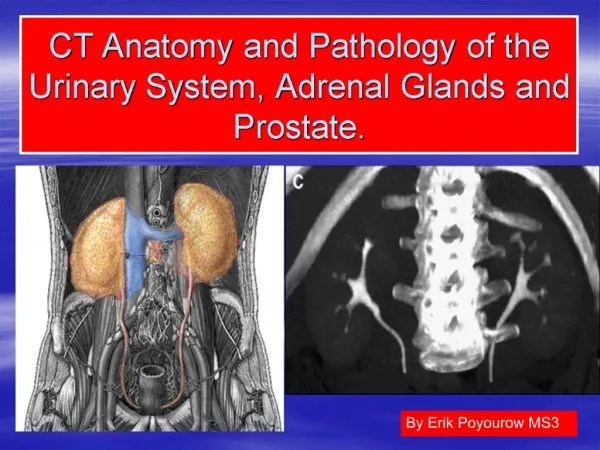
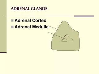
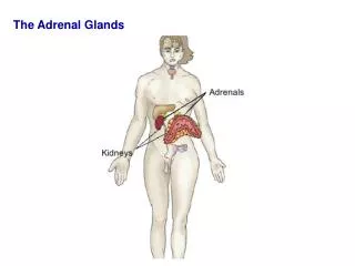
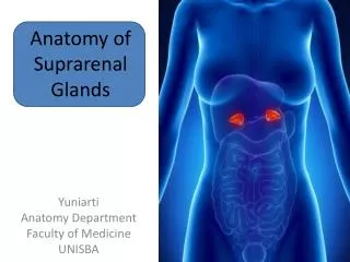
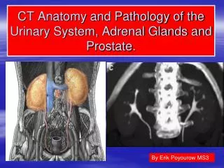
Anatomy of adrenal glands Location: the top of the kidney and weighting approximately 5 g each. Vascularization: a. suprarenalis superior (from a. phrenica inferior), a. suprarenalis media (from aorta abdominalis), a suprarenalis inferior (from a. renalis). Innervation: n. splanchnicus major (through plexus celiacus and plexus renalis), fibrae n. vagus and n. phrenicus. a. suprarenalis media a. supranenalis superior a. phrenica inferior Left adrenal gland Left adrenal vein Aorta a. suprarenalis inferior Vena cava inferior Left renal artery Right adrenal vein kidney Rena vein Right ovaria vein Left ovaria vein Ovaria arteries
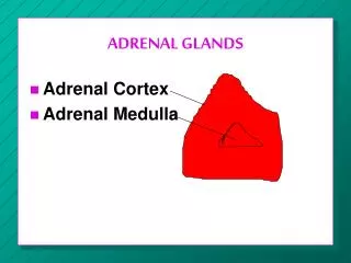
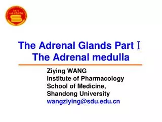
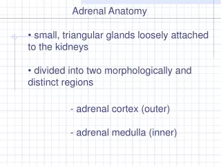
Anatomy of adrenal glands The adrenals are divided into: 1) outer area or cortex, which includes three zones: • Glomerular (glomerulosa) • Fascicular (fasciculata) • Reticular (reticularis) 2) inner area or medulla
The pituitary gland is the “master gland”, which lies in a bony structure, the sella turcica, located at the base of the skull. The gland is a small organ about I cm long; it weighs 500 mg and is divided into two parts, anterior (adenohypophysis) and posterior (neurohypophysis). The anterior pituitary secretes • corticotropin (ACTH) • Prolactin • Somatotropin (growth hormone (GH) • gonadotropins [follicle-stimulating (FSH) and luteinizing (LH) hormones] • thyrotropin (TSH) • melanocyte-stimulating hormones (MSH). In the nerve endings of the posterior pituitaryare stored • Vasopressin (antidiuretic hormone, ADH) • Oxytocin
The hypothalamusplays an important role in hormone regulation by secreting a series of small peptides which stimulate or inhibit the synthesis and release of hormones by the anterior pituitary • Realising Inhibiting • CRG • TRG • LGRG • FSRG • GRH - GIH (somatostatin) • PRG - PIF (dopamine) • MRG - MIH
Action of mineralocorticoids: regulation of electrolyte balance in the organism: • increasing the level of sodium (by sodium retention in distal nephron, colon, salivary gland) • decreasing the level of potassium (by excretion).
Action of glucocorticoids: • increasing of glycogen synthesis in liver and decreasing of glucose utilization by peripheral tissues, increasing gluconeogenesis; • increasing of protein synthesis in liver and decreasing of its synthesis in muscles and increasing of protein destruction in muscles; • increasing of lipolisis; • anti-inflammatory function and immunomodulation; • cardiovascular regulation (increasing of blood pressure).
Regulation • FEEDBACK: Hormone secretion → delivery to target cells → hormone recognition by receptors in target cells → biologic effect → hormone degradation → signal from target cells to stop further hormonesecretion
Regulation of secretion • glucocorticoids’ and androgens’ secretion is regulated by hypothalamic – pituitary system
mineralocorticoids’ secretion is regulated by the renin – angiotensin system, the level of Na+, K+ in blood, and to a lesser extent of ACTH
Action of catecholamines: • modulate vascular tone; • increase heart rate; • antagonize insulin action.
CHRONIC ADRENOCORTICAL INSUFFICIENCY. It is an insidious and usually progressive disease resulting from adrenocortical hypofunction.
Classification. • Primary adrenocortical insufficiency (Addison’s disease). • Secondary adrenocortical insufficiency .
Etiology of adrenal insufficiency: Primary: • autoimmune processes (50 – 65 %); • tuberculosis; • neoplasm, metastatic carcinoma; • inflammatory necrosis; • amyloidosis; heamochromatosis; • bilateral adrenal hemorrhage or infarction, intra – adrenal hemorrhage (Waterhouse – Friedrichsen syndrome following meningococcal septicemia); • bilateral adrenalectomy; Secondary: • hypothalamic or pituitary disease (primary injury of these organs leads to insufficiency of ACTH secretion that cause the two – side atrophy of adrenal glands); • glucocorticoid therapy.
Pathogenesis. Deficiency of adrenal hormones contributes to the hypotension and produces disturbances in carbohydrate, fat, and protein metabolism, and severe insulin sensitivity.
Symptoms and signs. Presentation may be acute and chronic. Frequently clinical signs of the primary chronic adrenocortical insufficiency are manifested in that time when adrenocortical tissue is destroyed on 70-90 %. The most common complaints are: • weakness, • malaise, • weight loss, • anorexia, • depression.
Objective examination: • Hyperpigmentation(in patients with primary adrenal insufficiency) is characterized by diffuse tanning of both exposed and nonexposed portions of the body, especially on pressure points (bony prominences), skin folds, scars, and extensor surfaces, black freckles over the forehead, face, neck, and shoulders; bluish – black discoloration of the areolas and the mucous membranes of the lips, mouth, rectum and vagina are common. After compensation hyperpigmentation will decrease. Patients in 15 – 20 % of cases may have areas of vitiligo(depigmentation) as the sign of autoimmune process.
Objective examination: • Hypotension or postural hypotension (88 – 90 %) with syncopal attacks can occur. • Tachycardia. • Weight loss (due to dyspeptic syndrome, true muscle tissue catabolism, dehydration). • Anorexia, nausea, vomiting, abdominal pain, diarrhea are often. Gastritis, ulcer disease can occur. • Decreased cold tolerance, with hypometabolism may be noted. • Sexual disorders. • Neurologic and psychiatric disorders: decreasing of the memory, mental activity, concentration of attention, depressions, hallucinations can occur due to chronic hypoglycemia which leads to changes of metabolism in brain tissue. • Hypoglycemia. There are three stages of severity: mild, moderate and severe.
Laboratory findings. • A low serum Na level and a high serum P level together with a characteristic clinical picture suggest the possibility of Addison’s disease. • Adrenal insufficiency can be specifically diagnosed by: • low levels of plasma glucocorticoids and mineralocorticoids, or urinary 17 – hydroxycorticosteroid (17 – OHCS) or 17 – ketogenic steroid (17 – KGS); • demonstrating failure to increase plasma cortisol levels, or urinary 17 – OHCS or 17 – KGS excretion, upon administration of ACTH(in patients with primary adrenal insufficiency, those with secondary adrenocortical insufficiency will have a significant increase in plasma cortisol or 24 - h urinary corticosteroid levels.) • To distinguish between primary and secondary adrenal insufficiency, me have to find the level ofplasma ACTH: primary shows increased, and secondary shows decreased level.
Instrumental findings. • The ECG may decreased voltage and prolonged P – R and Q – T intervals.
Treatment. • Etiologic: appropriate treatment of complicating infections (e.g., tuberculosis). • Pathogenic: • Diet (enough quantity of proteins, vitamins, salt and water). • Glucocorticoids (normally, glucocorticoids are secreted maximally in the early morning hours, little being secreted at night). Average dosage is: • cortisol: 20 – 25 mg daily; • prednisolone 5 – 7.5 mg daily; • hydrocortisone 30 – 40 mg orally daily. 2/3 of the dose can be given in the morning and 1/3 in the afternoon. Night doses should be avoided, as they may produce insomnia.
Mineraloocorticoids. DOCSA 5 mgorally daily should be used in patients with severe and moderate duration orfludrocortisone 0.1 – 0.2 mgorally once a day is recommended (this mineralocorticoid replaces aldosterone) • Intercurrent illnesses (e.g., infections) should be regarded as potentially serious and the patient should double his dosage until he is well. • If nausea or vomiting preclude oral therapy, medical attention should be sought immediately and parental therapy started.
Adrenal crisis - is a medical emergency caused by sudden marked insufficiency of adrenocortical hormones.
Precipitating factors. • stress (infection (especially with septicemia), trauma, surgery, prolonged fasting, salt loss due to excessive sweating during hot weather); • sudden withdrawal of adrenocortical hormone therapy in patients with chronic insufficiency.
Clinical features. An adrenal crisis is characterized by • profound asthenia, • severe pains in the abdomen, lower back or legs; • nausea, vomiting, diarrhea; • peripheral vascular collapse; • renal failure with azotemia. • Body temperature may be subnormal, through severe hyperthermia due to infection is often seen.
Treatment. Therapy should be instituted immediately once a provisional diagnosis of adrenocortical failure has been made. • Substitution therapy • Rehydration • Treatment of complications (hyperpyrexia, psychotic reactions).
Treatment. • hydrocortisone 100 – 150 mg as a water – soluble ester (usually the succinate or phosphate) is injected IV or acetate IM • followed by infusion of 1 L of 5 % glucose – in – saline solution containing 100 mg hydrocortisone ester given over 2 h. • Hydrocortisone acetate 50 – 75 mg IM each 4 – 6 h • Hydrocortisone therapy is given continuously to a total dosage in 24 h of 400 – 600 – 800 mg. • After stabilization of BP (>100 mm Hg) we decrease the dosage of hydrocortisone acetate to 25 - 50 mg IM to 2 - 4 times a week
Treatment. • Mineralocorticoids are not required when high – dose hydrocortisone is given • In a case of prominent hypotension DOKSA (5 mg), ftorhydrocortisone (cortinef 0,05 – 0,2 mg), fludrocortisone acetate (0.1 mg) have to be used • Total infusion of saline and 5 % glucose - 2,5 – 3,5 l during first day
Prognosis. With a substitution therapy, the prognosis is excellent and a patient with Addison’s disease should be able to lead a full life.
PHEOCHROMOCYTOMA. It is a tumor of chromaffin cells that secrete catecholamines • Rare: 5 per 100 000 hypertensives • Sporadic: 75 – 85 % • Hereditary: 15 – 25 % • Malignant: 3 – 36 % • Occurs equally in men and women • Peaks in 3rd – 5th decades
The term pheochromocytoma (phios means dusky, chroma means color, and cytoma means tumor) refers to the color the tumor cells acquire when stained with chromium salts
Etiologyis unknown • In about 80 – 90 % of cases, pheochromocytomas are found in the adrenal medulla, but may also be found in other tissues derived from neural crest cells (e.g., tumors may be found in the paraganglia of the sympathetic chain, retroperitoneally along the course of the aorta, in the carotid body, at the aortic bifurcation, in the GU system, in the brain, and in the dermoid cysts.
Classification. • Paroxysmal form (45 %). • Permanent form (50 %): • with crisis; • without crises. • Latent or silent form (nonsymptomatic).
Clinical features • hypertension, • Tachycardia, diaphoresis, postural hypotension, tachypnea, • flushing, • cold and clammy skin, severe headache, angina, palpitation, • visual disturbances, dyspnea, parasthesias • nausea, vomiting, epigastric pain, constipation or diarrhea and a sense of impending doom are common; some or all of these symptoms and signs may occur in any patient.
Symptoms “The 5 P’s” • Pressure increase (hypertension ) • Palpitation (tachycardia) • Perspiration • Pain (abrupt onset of throbbing headache, chest (angina), abdominal pain) • Pallor (due to vasoconstriction) cold and clammy skin
Other signs • postural hypotension or shock (epinephrine secretion) • tremor • visual disturbances • dyspnea • parasthesias • nausea, vomiting, • severe constipation or diarrhea and a sense of impending doom are common; • weight loss • anxiety • some or all of these symptoms and signs may occur in any patient.
Paroxysmal attacks may be • spontaneous or provoked by • palpation of a tumour, • postural changes, • abdominal compression or massage, • induction of anesthesia, • emotional trauma, • β – adrenergic blocking agents, • rarely, micturition.
Duration of hypertensive crisis is variable • from a seconds or few minutes to hours, • but 50 % of the paroxysms last less than 15 min. • Permanent form of the disease’s duration looks like malignant hypertension. • Nonsymptomatic form of the disease is rare.
Physical examination • hypertension • retinopathy • cardiomegaly • (but last two are often less extensive than might be expected for the degree of hypertension present)
Investigations • An increased 3-h (24-h) urinary excretion of epinephrine, norepinephrine and their metabolic products (VMA or metanephrines). • Increased plasma epinephrine, norepinephrine. • CT scanning, MRI of the abdomen for the localization of the tumor. • A scan with iodine I 131–labeled metaiodobenzylguanidine (MIBG) is useful for extra – adrenal tumors.
Treatment • Surgical removal of the tumor is the treatment of choice. 2. During crisis a combination of α- and β- adrenergic blocking agents • phentolamine (tropaphen) 2 - 4 mg every 5 - 10 min till stopping of the crisis, • phenoxybenzamine 10 – 20 mg 3 – 4 times daily, • propranolol 30 – 60 mg/day • and infusion of sodium nitroprusside.
Etiology of hypothalamo – pituitary disorders 1.Trauma 2. Infectious diseases: • acute (scarlet fever, influenza) • chronic (tuberculosis, malaria, toxoplasmosis) 3. Tumor or metastasis 4. Vascular damaging (thrombosis, thromboembolia) 5. Metabolic disorders (xanthomathosis) 6. Congenital pituitary hypo – or aplasia (syndrome of “empty sella turcica” 7. Genetic predisposition 8. Idiopathic
Pituitary tumors • Slow – growing (benign) • 10 – 15 % of all brain tumors • Clinical presentations: • Mass effect: • Visual deffects • Headaches • Seizures • Facial pain or numbness • Apoplexia • Nausea, vomiting 2. Hormonal effects (overproduction or deficiency)
Classificationof hypothalamo – pituitary disorders • Adenohypophysis disorders 1. Secretion of GH • overproduction: acromegaly, giantism • dificiency: pituitary dwarfism 2. Secretion of ACTH • overproduction: Cushing’s syndrome, hypothalamic syndrome 3. Secretion of Prolactin • overproduction: hyperprolactinemia, galactorhea-amenorhea 4. Secretion of TSH 5. Secretion of Gonadotropines: adiposogenital-dystrophy 6. Hypopituitarism 7. Hypothalamic obesity • Neurohypophysis disorders 1. Deficiency of vasopresin: diabetes insipidus 2. Inapropritiative secretion of vasopresin
GROWTH-HORMONE EXCESS(acromegaly and gigantism) Chronic, debilitating disorder resulting from excessive secretion of GH and resulting in production of insulin-like growth factor 1 (IGF-1), which lead to typical picture: gigantismbefore puberty and to acromegaly after puberty
Etiology • Adenoma (acidophilic or chromophobe) of the pituitary • Ectopic GH-producing tumors (cancer of lungs, mamma glands, pancreatic gland) • Regulatory hypothalamus disturbances (overproduction of somatoliberin or deficiency of somatostatin) • Ectopic GRH-producing tumors (carcinoid tumor of intestine, lungs)
Clinical manifestations of gigantism • Mass effects • Overproduction of GH • height more than 190 cm in women and 200 cm in men • Hypersecretion of GH prior to closure of epiphysis leads to proportional growth of bones; both length and width of bones are increased • May be blood hypertension, mycardial dystrophy, atrial fibrillation
Acromegaly • Named “acromegaly” by Pierre Marie in 1886 from Greek akron (extremities) and megas (large) • Rare disease • 3 – 4 new cases per million population per year • Affects both sexes equally • Peak ages 40 – 60 years • Mortality 10 times higher than in control group mostly due to cardiovascular disorders
Clinical manifestations of acromegaly • Syndromes connected with influence of GH on organs and tissues: • periostal overgrowth: • overgrowth of the mandible leads to protrusion of the jaw (prognatism) • there is an overbite and the teeth become separated (diastema).