Download
1 / 64
650 likes | 789 Views
The Knee. Lesson one – Bone, ligaments and meniscus. One of the most complex joints in body Considered a hinge joint Flexion and extension Does have some rotational movement Internal and external Muscles and ligaments provide majority of stability. Bones. Femur Major reference points

E N D
The Knee Lesson one – Bone, ligaments and meniscus
One of the most complex joints in body • Considered a hinge joint • Flexion and extension • Does have some rotational movement • Internal and external • Muscles and ligaments provide majority of stability
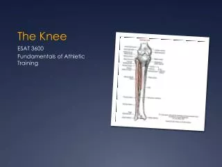
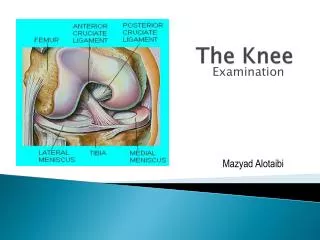
Bones • Femur • Major reference points • Lateral and medial (femoral) condyles • Tibia • Lateral and medial (tibial) condyles • Tibial plateau • Tibial tuberosity • Patellar tendon insertion • Gerdy’s tubricle • IT band insertion
Bones • Fibula • Non weight bearing bone • Used more muscle and ligament attachments • Patella • Largest sesmoid bone in the body • Located in the quadriceps tendon • Tracks with the concavity formed by the femoral condyles • Improves the force action of the quad tendon
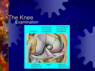
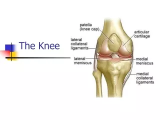
Meniscus • Fibrocartilage that lies on the tibial plateau • It helps improve the continuity between the femur and tibia. • It cushions the area • Helps the synovial fluid be spread out in the joint capsule • Has poor blood supply • Thus it heals poorly • Only the outer edges have any sort of blood supply.
Mensicus cont. • Medial portion is C-shaped and attached to the tibial collateral ligament thus is more stable • This causes it to be injured more often than lateral • Lateral portion is O-shaped and is largely unattached • Three Ligaments help them stay in place; transverse ligament, ligament of Wrisberg, and coronary ligament
Collateral Ligaments • Tibial collateral (MCL) • Supports medial stress on the knee • Secondary restraint is anterior displacement • Originates - medial epicondyle of femur • Inserts • Superficial – on medial tibial condyle • Deep – blends with capsule and semimembranosus and attaches to medial meniscus
Collateral ligaments • Fibular collateral ligament (LCL) • Gives lateral support against varus stress to the knee • Secondary restraint is ant/post displacement • Originates - lateral femoral epicondyle • Inserts - the head of the fibula
Cruciate ligaments • Anterior cruciate (ACL) • Keeps femur from moving posterior and the tibia from moving anterior • Protects against abnormal internal rotation • Originates - on anterior tibia • Inserts - inner surface of lateral femoral condyle
Cruciate ligaments • Posterior cruciate (PCL) • Prevents hyperextension of knee • Prevents from abnormal forward sliding of the femur • Protects against abnormal external rotation • Originates – posterior tibia • Inserts - medial femoral condyle (anterior lateral surface)
Deep medial capsular ligaments • Transverse ligament • Joins the anterior portion of the medial and lateral meniscus • Ligament of Wrisberg • Holds the posterior lateral meniscus • Coronary ligaments • Medial and lateral • Part of joint capsule that holds the medial and lateral meniscus to the medial and lateral tibial condyles, respectively
Other structure • Bursas • Thin pads that are located in high friction areas • Located in • Superior knee • Pre patella • Inferior knee • Posterior knee (2)
Fat pad • Located under the patella tendon • Reduces friction and gives cushion to area
Popliteal fossa • The opening in the back of the knee; formed from the tendons of the hamstrings and gastrocnemius.
Common Peroneal nerve • Wraps around the head of the fibula and traverses down the lateral leg • Tibial nerve
The Knee Lesson two - Muscles
Quadriceps • Quadriceps consist of four muscles covering the anterior portion of the thigh. • The four muscles blend together on the top of the patella and form the quad tendon. This encompasses the patella. • This forms into the patella tendon/ligament which inserts on the tibial tuberosity
Rectus femorus (1) • Two joint muscle – flexes hip and extends knee • Meaning it crosses two joints and thus helps with two motions • Originates on Anterior inferior iliac spine (AIIS) of Ilium
Vastus lateralis (2) • Originates on outer femur below greater tuberosity • Vastus Medialis (4) • Originates -whole length of inner femur • Vastus Intermedias • Originates on upper two-thirds of outer femur • Lies under Rectus Femorus
Hamstrings • All muscles originate on the ischial tuberosity • The muscles cover the whole posterior side of the thigh • All of the muscles are two joint muscle. • They all help extend hip and flex knee
Hamstrings cont. • Biceps femorus (1,2) • Inserts on Fibular head • External rotation • Semitendinosus (3) • Inserts on anterior medial tibial condyle • Helps form the pes anserine (gracilis and sartorius) • Internal rotation • Semimembranosus (4) • Inserts on posterior surface of medial tibial condyle • Internal rotation
Adductors • Adductor Brevis • Origin– front of pubis • Insertion – lesser trochanter • Action – adduction of hip • Rotation outward as it adducts the hip
Adductors cont. • Adductor longus • Origin – front of pubis • Insertion – middle third of the linea aspera • Action – hip adduction • Assists in hip flexion
Adductors cont. • Adductor magnus • Origin – Edge of entire pubis and the ischium and ischium tuberosity • Insertion - whole length of linea aspera and inner condyloid ridge • Action – hip adduction and rotation outward with hip adduction
Adductors cont. • Gracilias • Origin - Inner edge of descending ramus of the pubis • Inserts - anterior medial surface of tibia below condyle • Part of Pes ansernine muscle group (semitendinosus and sartorius) • Action - two joint muscle • Adduction of hip • Flexion of knee • Inward rotation of the hip
Other muscles…. • Sartorius • Origin – notch between ASIS and AIIS • Insertion – anterior medial tibial condyle • Action – weak hip flexor and knee flexor (two joint muscle).
Iliopsoas • Origin - ilium, sacrum, and sides of the the 12th thoracic vertabrae and all the lumbar vertabrae. • Insert – Lesser trochanter of the femur and part of shaft distal of point. • Action – flexion of the hip
Popliteus • Origin – posterior surface of lateral femoral condyle • Insertion – upper posterior-medial surface of the tibia • Action – flexion of the knee
Tensor Fascia latae • Origin – anterior iliac crest and anterior surface of ilium • Insertion – IT band formation 1/4 way down lateral thigh • Action – weak hip flexion, horizontal abduction, mild rotation of the hip. • Forms IT band that courses down side of leg and inserts on a point on tibia called Gerdy’s tubercle
The Knee Lesson three - injuries
Anterior Cruciate injury • Common etiology • Externally rotated and knee put into valgus stress • Blow or excessive shifting force • Symptoms • Hear a pop or snap • Feeling of tibia shifting forward • Rapid effusion
ACL cont. • Tests • Anterior Drawer and Lachman’s • Treatment • RICE • Bracing and crutches • Referral to Orthopedic • additional info • Hamstrings support ACL • Not uncommon to injure other structures
Posterior Cruciate • Etiology • Knee slightly flexed and anterior force on shin • falling on bent knee • Symptoms • similar to ACL • Tests • Posterior drawer • Sag test • additional info • not as serious as ACL • quads support PCL • surgery not always done
Tibial collateral ligament (MCL) sprain • Etiology • Valgus stress • symptoms • pain over medial knee • inability to walk • swelling over medial knee • laxity with valgus test
MCL cont. • Tests • Valgus stress test • 0 degrees – deep MCL • 30 degrees – superficial MCL • Degrees of sprain • 1st degree – slight laxity no pain • 2nd degree – laxity but still have end point • 3rd degree – opens up completely • Treatment • RICE • Place in brace • Doctor referral
Fibular collateral ligament (LCL) sprain • Etiology • Varus stress • Twisting • Symptoms • Similar to MCL • Tests • Varus stress • Treatment • RICE • Referral to doctor
Meniscus • Etiology • Twisting action • Valgus stress • Symptoms • Popping and clicking over joint line • Pain over joint line • Giving away • Locking of knee • Slight swelling over knee
Meniscus cont. • Tests • McMurray’s • external rotation – medial • internal rotation – lateral • Apley’s compression/distraction test • Additional info • Types of injuries • longitudinal or bucket handle • posterior horn tear • parrot beak • Surgeries • meniscal repair • 3-4 months rehab • menisectomy • snip out torn parts • 4-8 weeks rehab