Download
1 / 45
450 likes | 456 Views
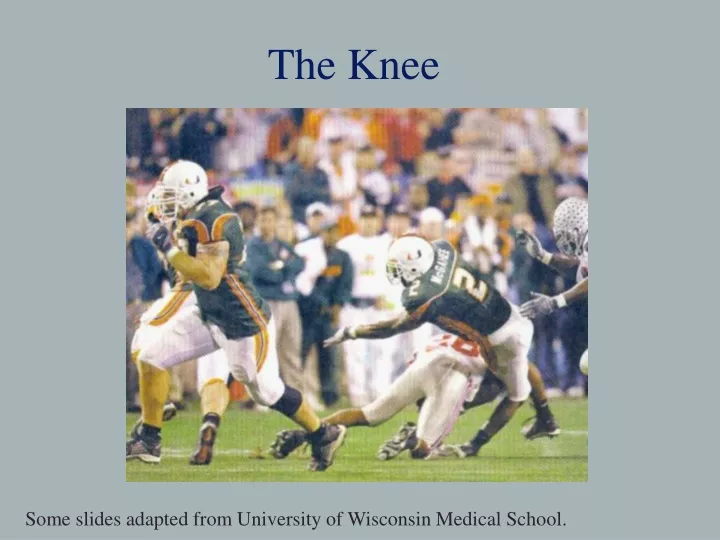
The Knee. Some slides adapted from University of Wisconsin Medical School. The Knee. One of the most complex joints Provides stability in weight bearing and locomotion Very vulnerable – especially medially and laterally Muscles and ligaments provide most of the stability.

E N D
The Knee Some slides adapted from University of Wisconsin Medical School.
The Knee • One of the most complex joints • Provides stability in weight bearing and locomotion • Very vulnerable – especially medially and laterally • Muscles and ligaments provide most of the stability
Instability - Example Patellar dislocation http://www.carletonsportsmed.com/Libraria_medicus/PF_patella_dislocation.JPG
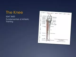
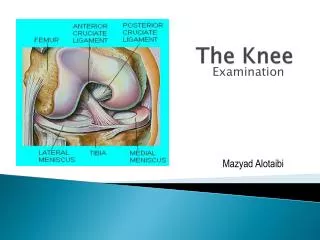
Bony Anatomy – 4 bones Femur Patella Tibia Fibula
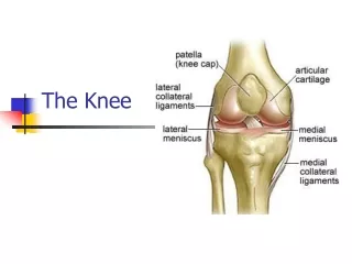
Bony Anatomy • Femur: Longest Bone in Body • Tibia: WB bone of lower extremity • Fibula: Site of Muscle Attachment • Patella: Sesamoid Bone • Floating bone • A bone that develops within a tendon
Knee Skeletal Lateral Condyle Head of Fibula Femoral Groove Gerdy’s Tubercle Tibial Tuberosity Pes Anserine
Knee Menisci • 2 oval shaped (semilunar) fibrocartilages • Provides cushion • Avascular(poor blood supply) = decreased healing • Medial – “C” shaped • Lateral – “O” shaped
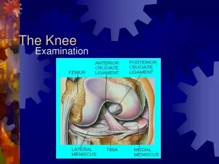
Menisci Medial Meniscus Lateral Meniscus PCL ACL
4 Stabilizing Ligaments Posterior Cruciate • 2 cruciate ligaments • ACL/PCL • 2 collateral ligaments • MCL/LCL Anterior Cruciate Medial Collateral Lateral Collateral
Anterior Cruciate Ligament (ACL) • Extends from tibia posteriorly and laterally to femur • Front of tibia to back of femur • Prevents anterior movement of tibia • Stabilizes against tibial rotation • Main stabilizer
ACL • Torn during cutting motions • Foot planted and knee rotates • More commonly torn in girls • Less muscle, hormones, Q- angle • Surgery • Cadaver graph, patellar tendon, hamstring tendon • About 6-9 months to return to activity
Posterior Cruciate Ligament (PCL) • Extends antiorly and medially from tibia to posterior femur • Prevents tibia from posterior translation • Prevents hyperextension
Medial Collateral LigamentMCL • Medial side • Thick Band of Tissue • Tibia Femur • Resists Valgus Force
Valgus • Outside to Inside Force • MCL resists this force • Occurs in FRONTAL PLANE
Lateral Collateral Ligament LCL • Lateral side • Narrow cord-like band of tissue • Connects femur to • head of fibula • Resists Varus Force
Varus • Inside to Outside Force • LCL resists this force • FRONTAL PLANE
Collateral Ligament Ruptures • 3 degrees of sprains (ligament damage) • Complete tear = 3rd degree sprain
Ruptured Patellar Tendon 3rd degree Strain = muscle/tendon injury
Lab Activity • Partner up • Get a marker • Identify structures of the knee • Patella • Head of fibula • Tibial tuberosity • Pes Anserine • Gerdy’s Tuburcle • MCL • LCL • Medial Joint Line • Lateral Joint Line • Patellar Tendon
Surface Anatomy - Anterior, Extended* Patella Indented Hollow
Surface Anatomy - Anterior, Flexed Patella Tibial Tuberosity Head Of Fibula
Palpation – Anterior* Patella: Lateral and Medial Patellar Facets Superior And Inferior Patellar Facets Medial Fat Pat Lateral Fat Pad Patellar Tendon**
Surface Anatomy - Medial Patella Tibial Tuberosity Medial Femoral Condyle Joint Line Medial Tibial Condyle
Palpation - Medial Medial Collateral Ligament (MCL)* Pes anserine bursa** Medial joint line
Surface Anatomy – Lateral Patella Quadriceps Tibial Tuberosity Head Of Fibula
Palpation – Lateral* Lateral Collateral Ligament (LCL)** Lateral joint line
You should have the following drawn on your partner’s knee • Patella • Head of fibula • Tibial tuberosity • Medial joint line • Lateral joint line • Patellar tendon • MCL • LCL • Pes Anserine • Gerdy’s tubercule
Quadriceps and Patellar Tendons • Quadriceps Tendon • All 4 muscles come together at patella • Patellar Tendon • From inferior patella to tibialtuberosity
Quadriceps • Anterior Thigh Musculature • Four Muscles: • Rectus Femoris • Vastus Lateralis • Vastus Medialis • Vastus Intermedius • Extend the Knee
Rectus Femoris • 2 Joint Muscle • Crosses hip and knee • Flexes Hip • Extend the knee • Converges with rest of quadriceps muscles at tibial tubercle
Hamstrings • Three Muscles • Semimembranosus • Semitendinosus • Biceps Femoris • Common Origin the ischial tuberosity • Flex the Knee