Download
1 / 45
740 likes | 1.77k Views
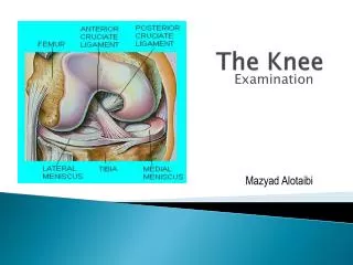
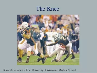
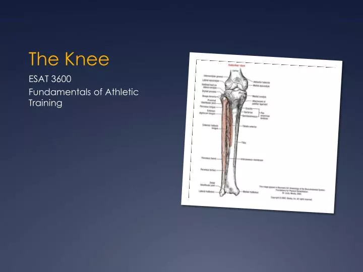
The Knee. ESAT 3600 Fundamentals of Athletic Training. Knee Complex. 2 articulations Tibiofemoral (knee joint ) Medial and lateral Patellofemoral. Tibiofemoral Joint. Articulation of the femur and tibia Medial and lateral articulating surfaces Femur has convex surfaces

E N D
The Knee ESAT 3600 Fundamentals of Athletic Training
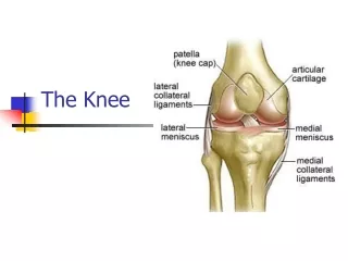
Knee Complex • 2 articulations • Tibiofemoral (knee joint) • Medial and lateral • Patellofemoral
Tibiofemoral Joint • Articulation of the femur and tibia • Medial and lateral articulating surfaces • Femur has convex surfaces • Tibia has concave surfaces
Patellofemoral Joint • Articulation of patella with femur • Patella serves as pulley mechanism for quadriceps muscles • PFPS • Chondromalacia
Bony Landmarks (Femur) • Lateral condyle • Lateral epicondyle • Medial condyle • Medial epicondyle • Adductor tubercle • Poplitealfossa • Intercondylar notch • Patellar facet
Bony Landmarks (Tibia) • Tibialtuberosity • PesAnserines • Gerdy’s Tubercle
Pes Anserines • Point of insertion of sartorius, gracilis, and semitendinosus
Gerdy’s Tubercle • Small prominence on anterior aspect of lateral condyle of tibia • Insertion of IT band
Superior View of Tibia • Intercondylarfossa • Posterior • Anterior • Intercondylar eminence • Medial articular surface • Lateral articular surface
Patella • Base • Apex • Lateral border • Medial border • Lateral articulating surface • Medial articulating surface
Knee Movements • Flexion • Extension • Medial rotation • Lateral rotation
Arthrokinematics of Tibiofemoral Extension • Matter of perspective • Tibia moving on fixed femur • Femur moving on fixed tibia
Screw-Home Mechanism • 3 factors • Shape of medial condyle • Passive tension of ACL • Lateral pull of quadriceps • Also a matter of perspective • External rotation of tibia on femur • Internal rotation of femur on fixed tibia
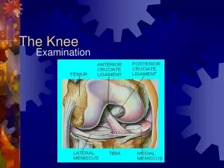
Knee Stability • Bony stability is extremely weak • Ligaments and cartilage provide most stability
Menisci and Attachment Sites • Medial • C-shaped • Lateral • Incomplete O
Ligament of Wrisberg • Lateral meniscus to posterior medial condyle
4 Main Functions of Menisci • Maintain congruence between articular surfaces in all positions of the joint • Provide shock absorption in the joint • Maintain circulation of synovial fluid through articular cartilages • Help bring about normal movement between the articular surfaces
Meniscal Injury • Tearing attachments of the menisci to the tibial table and joint capsule • Crushing the menisci between the femoral and tibial condyles, produces circular (bucket handle) and radial tears
Joint Capsule • Common capsule for tibiofemoral and patellofemoral joints • Anterior part folds upward during extension • Posterior part folds downward during flexion
Knee Ligaments • Lateral collateral • Attached superiorly to lateral epicondyle, inferiorly to head of fibula • Medial collateral • Attached superiorly to medial epicondyle, inferiorly to medial aspect of tibia below tibial condyle
Knee Ligaments • Anterior cruciate • Distal attachment – posterior aspect of anterior intercondylar area of tibia • Proximal attachment – posterior medial aspect of lateral femoral condyle
ACL • Anteriomedial band • Tight in flexion • Posteriolateral band • Tight in extension • Both are tight in extension • PL band is more tight
Knee Ligaments • Posterior cruciate • Distal attachment – posterior aspect of posterior intercondylar area of tibia • Proximal attachment – anterior inferior lateral aspect of medial femoral condyle
Role of Cruciate Ligaments • Bring about normal movement between articular surfaces • ACL – prevents anterior displacement of tibia relative to femur • Prevents medial rotation • PCL – prevents posterior displacement of tibia relative to femur
Posterior Knee Ligaments • Oblique popliteal ligament • Runs from posterior aspect of the lateral condyle of femur to posterior edge of medial condyle of the tibia • Protects against hyperextension
Posterior Knee Ligaments • Arcute ligament • Runs from the posterior aspect of the lateral condyle to the posterior surface of capsular ligament • Protects against hyperextension
Ligamentous Stability in General • Not constant throughout ROM • Knee extended most stable • Knee flexed least stable
Q Angle • Pull angle of the quadriceps • 8 – 17 degrees is normal • Increased angles associated with patellofemoral problems
Knee Alignments • Genuvalgum • Genuvarum • Genurecurvatum
Knee Function • Dual role of mobility and stability • Mobile enough to allow movement • Stabile enough to absorb forces • Gait and hamstrings
Muscles Covered With the Hip • Sartorius • Rectus Femoris • Tensor FaciaLata • Gracilis • Biceps Femoris • Semitendinosis • Semimembranosus • See book for review
Vastus Medialis • O: lower ½ of intertrochanteric line, medial lip of linea aspera, upper part of medial supracondylar line, medial intermuscular septum, tendon of adductor magnus and longus • I: medial border of patella , through patellar ligament to tibial tuberosity • A: extends the leg at the knee and draws patella medially
Vastus Intermedius • O: proximal 2/3 of the anterolateral surface of femur, lower ½ of the linea aspera, upper part of lateral intermuscular septum • I: by tendons of rectus femoris and vasti muscles into superior border of patella, through patellar ligament to tibial tuberosity • A: extends leg at knee
Vastus Lateralis • O: upper part of intertrochanteric line, anterior and lower borders of greater trochanter, lateral lip of gluteal tuberosity, upper half of linea aspera, lateral intermuscular septum, and tendon of the gluteus maximus • I: lateral border of patella and through patellar ligament to tibial tuberosity • A: Extends leg at knee and draws patella leterally
Popliteus • O: lateral condyle of femur, outer margin of lateral meniscus, arcuate popliteal ligament and capsule of knee joint • I: posterior surface of tibia above soleal line • A: rotates the tibia medially on the femur, or the femur laterally on the tibia (depends on the one fixed)
Gastrocnemius • O: lateral condyle and posterior surface of femur, capsule of knee joint. Medial condyle and adjacent part of femur • I: posterior surface of calcaneus by means of achilles tendon • A: flexes leg at the knee, plantar flexion and inversion of foot
Muscle Action Around the Knee • All create stability of joint • Hamstrings help prevent ATD • 2-Joint arrangement provides efficiency of movement • 2-joint arrangement can lead to problems • Passive insufficiency • Active insufficiency
Knee Instability • Knee is prone to instability and injury • Continuous stress • Beyond limit of ROM • Rotation with foot fixed • Most rotation with knee flexed • Mobile adapter for twisting/turning
Deep Squats • Safety dependent on how performed • Ability of knee to absorb forces dependent on: • Speed of descent • Size of calves and thighs • Strength of muscles controlling movement
Deep Squats • Dangerous when center of knee rotation is changed because of calf and thigh tissues pressing together • Lean forward at trunk to adjust center of gravity towards knees • Muscle strength