Download
1 / 32
320 likes | 328 Views
Microscopy. Cell Theory. Living organisms either multicellular (made up of many cells) or unicellular (exist as single cells) Metabolic processes take place within cells New cells are derived from existing cells

E N D
Cell Theory • Living organisms either multicellular (made up of many cells) or unicellular (exist as single cells) • Metabolic processes take place within cells • New cells are derived from existing cells • The genetic material of the organism is passed from parent cells to daughter cells • A cell is the smallest unit of an organism capable of surviving independently Zooming into DNA
Check that lowest power of objective lens is clicked into place • Clip slide onto stage • Ensure that stage is closest to the lens • Look at the stage from the side and position slide so that the specimen is beneath the lens • Look down the eyepiece and carefully turn coarse focus until specimen comes into view • Adjust illumination • Use fine focus to adjust if necessary • To view with higher power make sure that the area of interest is in the centre of the field of view and then rotate objective lenses Microscopes through history Parts of the microscope quiz
Human tissue culture cell – false colour Prostate cancer cell Electron beam Specimen Copper grid Screen
Fly foot Butterfly proboscis Wellcome Institute SEM How the SEM works SEM Images
size of image I magnification = actual size M A size of image actual size = magnification Magnification • The magnification of an object is how many times bigger the image is when compared to the original object
size of image magnification = actual size Microscopic Units • 1 millimetre = 1000 micrometres (µm) • 1 micrometre = 1000 nanometres (nm) Image Actual = 2 mm = 8 mm = x 4
6000 = = x 300 20 6000 = = 40 µm 150 size of image magnification = actual size size of image actual size = magnification Image = 6mm ; Actual size = 20µm Magnification = ? Hint: keep the units the same ! Image = 6mm Magnification = x 150 Actual size = ?
X 6 Resolution Increasing magnification doesn’t always increase the resolution
Resolution is the ability to distinguish between two points – a lack of resolution means that the two points would appear as one • The resolving power of a microscope is related to the form of radiation being used to see the specimen • Using a light microscope objects 0.2µmor more apart can be seen separately
largest algal cell leaf (thickness) page plant cell animal cell large bacterial cell mitochondrion cell membrane 50 000 µm (50mm) 500 µm (0.5mm) 100 µm (0.1mm) 40 µm (0.04mm) 20 µm (0.02mm) 5 µm (0.005mm) 1 µm (0.001mm) 7nm (0.007µm) Size is everything… From coffee bean to carbon atom
=1mm =1000µm =0.5mm =500µm =0.1mm =100µm =0.04mm =40µm =0.02mm =20µm =0.005mm =5µm
Microscope drawingTitle/ Aspect(T.S. L.S ) Mag • Low Power Plans of tissues • Do not draw individual cells unless specifically told to do so. • No shading. • Ruled lines which do not cross. • Medium and High Power drawings • Show individual cells. • Only label cell organelles that are visible e.g. Cell wall, chromosomes, cytoplasm. • No shading etc. as above.
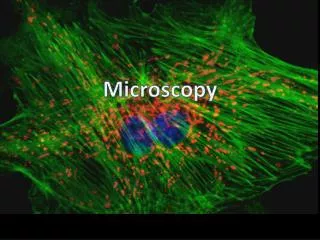
Gorgeous photomicrographs Nikon Small World Royal Microscopical Society; Biannual competition