Download
1 / 61
710 likes | 1.18k Views
MICROSCOPY. "micro" (small) "scopeo" (to watch). THE RELATIVE SIZES OF MOLECULES, CELLS AND ORGANISMS. THE RELATIVE SIZES OF MOLECULES, CELLS AND ORGANISMS. MICROSCOPY. MICROSCOPY. 1590. 200 9. THE LIGHT.

E N D
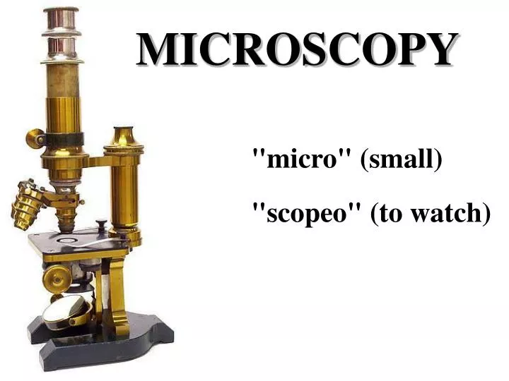
MICROSCOPY "micro" (small) "scopeo" (to watch)
MICROSCOPY 1590 2009
THE LIGHT Light: electromagnetic radiation of a wavelength that is visible to the human eye (about 400–700 nm). Light can exhibit properties of both waves and particles (photons). This property is referred to as wave–particle duality. Monochromatic light: light ray possessing one single wavelength Complex light: mixture of light rays with more different wavelengths.
THE LIGHT Characteristic parameters of light waves: Wavelength: the distance between repeating units of a propagating wave of a given frequency.(λ, expressed in nm). Frequency: The number of oscillations within a minute. Amplitude: distance from the center y position to the peak
MAGNIFICATION Total visual magnification of the microscope is derived by multiplying the magnification values of the objective and the eyepiece. Ocular: 5-30 x magnification Objective: 4-100 x magnification Maximum magnification: 3000X Can we apply the maximal magnification?
~0.25 mm The resolution Resolution: the shortest distance between two points on a specimen that can still be distinguished by the observer or camera system as separate entities. The resolution of our optical equipment is better as closer points can be seen as separate ones.
objective object The resolution of a light microscope The resolution of the microscope, () : () = 2 A = n·sinα The letter n is the refraction index of the media between the cover slip and the objective (air n=1, distilled water n=1.33, cedar oil n=1.51). α labels the angle closed by the main optical axis and the outermost light beam (half angle of the objective)
Possibilities of resolution improvement • Reduction of the numerator, i.e. application of light beam with a shorter wavelength. • With UV light the resolution can be reduced to 0.1 μm, but special quartz lenses and UV-light detector are needed, therefore the light microscope with UV light source is only a theoretical possibility.
Possibilities of resolution improvement (ii) To increase the value of the numeric aperture (A = n·sinα). Increasing n (refraction index): The resolution of the microscope can be enhanced by dropping a solution with higher refractive index between the front lens and the coverslip. Those lenses (mainly objectives with 100x magnification) are named as immersion objectives. The immersion liquid mentioned above is cedar oil, thus these lenses are objectives with oil immersion (they are labeled with HI). For the WI labeled objectives distilled water is the liquid which should be used. Increasing α: The half angle of a lens can be increased only until 72°, since at larger angle than this the light beams became totally reflected.
MICROSCOPIC MEASUREMENT objective micrometer ocular micrometer
MICROSCOPIC MEASUREMENT A piece aof hair objective micrometer scale ocular micrometer scale ocular micrometer scale
ADVANCED MICROSCOPY • Phase contrast microscopy • Fluorescence microscopy • Laser scanning confocal microscopy • Electron microscopy
Phase contrast microscopy A large spectrum of living biological specimens are virtually transparent when observed in the optical microscope under brightfield illumination. Phase contrast microscopy provides an excellent method of improving contrast in unstained biological specimens without significant loss in resolution, and is widely utilized to examine dynamic events in living cells. Fritz Zernike received a Nobel prize in 1953 for his discovery of phase contrast. In a phase-contrast microscope, the annular rings in the objective lens and the condenser separate the light. The light that passes through the central part of the light path is recombined with the light that travels around the periphery of the specimen. The interference produced by these two paths produces images in which the dense structures appear darker than the background.
Fluorescence microscopy/ Laser scanning confocal microscopy
Fertilization: Sperm aster, male and female pronuclei MT,DNA,CS
Transfected tissue culture cells DNAmicrotubules F-actin
Microscopy: the past and present intestine cross section, haematoxilin/eosin staining intestine cross section, multicolor fluorescence image
Microscopy: the past and present testis cross section, haematoxilin/eosin staining testis cross section, multicolor fluorescence image
Fluorescence Fluorescence - The process by which a suitable atom or molecule, which is transiently excited by absorption of external radiation at the proper energy level (usually ultraviolet or visible light), releases the absorbed energy as a photon having a wavelength longer than the absorbed energy. The fluorescence excitation and emission processes usually occur in less than a nanosecond.
Fluorescence Microscopy Fluorescence Microscopyis the most rapidly expanding microscopy technique employed today, both in the medical and biological sciences. When coupled to the optical microscope, fluorescence enables investigators to study a wide spectrum of phenomena in cellular biology. Foremost is the analysis of intracellular distribution of specific macromolecules in sub-cellular assemblies, such as the nucleus, membranes, cytoskeletal filaments, mitochondria, Golgi apparatus, and endoplasmic reticulum. In addition to steady state observations of cellular anatomy, fluorescence is also useful to probe intracellular dynamics and the interactions between various macromolecules, including diffusion, binding constants, enzymatic reaction rates, and a variety of reaction mechanisms, in time-resolved measurements. For example, fluorescent probes have been employed to monitor intracellular pH and the localized concentration of important ions.
Fluorescence Filter Cube Structure Exciter filter: permits only selected wavelengths from the illuminator to pass through on the way toward the specimen. Barrier filter: blocks the excitation wavelengths and permit only selected emission wavelengths to pass toward the eye or other detector. Dichromatic beamsplitter (dichroic mirror): reflects excitation wavelengths and passes emission wavelengths.
Fluorescence Microscope Structure Microscopes with an inverted-style frame are designed primarily for tissue culture applications and are capable of producing fluorescence illumination through an episcopic and optical pathway. Epi-illuminators usually consist of a mercury or xenon lamphouse (or laser system) stationed in a port at the rear of the microscope frame. Fluorescence illumination from the arc lamp passes through a collector lens and into a cubethat contains a set of interference filters, including a dichroic mirror, barrier filter, and excitation filter. Light reflected from the dichroic mirror is restricted in wavelength by the excitation filter and enters the objective (now acting as a condenser) to bathe the specimen with a cone of illumination whose size and shape is determined by the objective numerical aperture. Secondary fluorescence, emitted by the specimen, returns through the objective, dichroic mirror and barrier filter before being routed through the microscope optical train. The microscope presented above contains a trinocular observation tube that is equipped with a port and extension tube for mounting a traditional or CCD camera system (a Peltier-cooled CCD camera is illustrated). Another port, located near the base at the front of the microscope, can also serve as an attachment point for a camera system (a traditional 35-millimeter camera is shown in the figure). In the figure presented above, wide-spectrum fluorescence illumination is filtered to produce a narrow bandwidth of green excitation wavelengths, which are capable of exciting specific fluorophores in the specimen. Secondary fluorescence (red light) passes back through the objective and is distributed throughout the microscope optical system. Transmitted illumination is provided by a tungsten-halogen lamphouse that is positioned on the microscope pillar, above the stage. Light from the lamphouse passes through a collector lens, a series of filters, and the field diaphragm before entering the condenser front aperture. After being focused by the condenser lens elements, transmitted illumination is projected onto the specimen, which is placed on the stage. The light that is diffracted, refracted, and not absorbed by the specimen continues through the objective and into the microscope optical train where it can be directed to the eyepieces or to a camera system.
Fluorochromes Fluorochrome - A natural or synthetic dye or molecule that is capable of exhibiting fluorescence. Fluorochromes (also termed fluorescent molecules, probes, or fluorescent dyes) are usually polynuclear heterocyclic molecules containing nitrogen, sulfur, and/or oxygen with delocalized electron systems and reactive moieties that enable the compounds to be attached to a biological species.
Fluorochromes Bovine pulmonary artery endothelial cells visualized using components of the SelectFX Nuclear Labeling Kit and Alexa Fluor phalloidin conjugates. Nuclei and F-actin were stained, respectively, with (top left) DAPI and Alexa Fluor 680 phalloidin, (top right) SYTOX Green dye and Alexa Fluor 568 phalloidin, (bottom left) 7-AAD and Alexa Fluor 488 phalloidin, and (bottom right) TO-PRO-3 dye and Alexa Fluor 350 phalloidin.
Organelle/DNA/Ca2+ probes Organelle probes: a fluorochrome nucleus attached to a target-specific moiety that assists in localizing the fluorophore Mitochondrium: MitoTracker and MitoFluor Lysosome: LysoTracker and LysoSensor Golgi apparatus: BODIPY Endoplasmic reticulum: DiOC, Blue-White DPX DNA: Acridine orange, Propidium iodide, DAPI, Hoechst Ca2+: fura-2 and indo-1, fluo-3, fura red
Immunofluorescence Microscopy A targeted molecular species (protein, nucleic acid, membrane, etc.) in a specimen is labeled with a highly specific fluorescent antibody. After the labeled antibodies have been excited by a selected region of wavelengths, secondary fluorescence emission is gathered by the objective to form an image of the specimen. Antibodies are labeled either by coupling directly with a fluorochrome (fluorescent dye; termed direct immunofluorescence), or with a second fluorescent antibody that recognizes epitopes on the primary antibody (indirect immunofluorescence).
Immunofluorescence Microscopy MTDNSCS
Fluorescent Proteins Green Fluorescent Protein (GFP) - A naturally occurring protein fluorescent probe derived from the jellyfish Aequorea victoria, which is commonly employed to determine the location, concentration, interactions, and dynamics of a target protein in living cells and tissues. The excitation and emission spectra of enhanced GFP (a genetic derivative) have maxima at 489 nanometers and 508 nanometers, respectively. In order to incorporate the GFP (or any of its genetic derivatives) into a cell, the DNA sequence for the gene is ligated to the DNA encoding the protein of interest. After cultured cells have been transfected with the modified DNA, they are able to express chimeric fluorescent proteins for observation in the microscope. There are genetically modified variants of GFP such as blue fluorescent protein (BFP), cyan fluorescent protein (CFP), yellow fluorescent protein (YFP) DsRed fluorescent protein
Fluorescent Proteins Tubulin-GFP
GFP GFP GFP GFP-FUSION PROTEINS gene of interest plasmid with GFP gene fusion protein emits green light upon excitation by blue light, intracellular localization determined transfection GFP transcription, translation
Confocal Laser Scanning Microscopy Confocal Laser Scanning Microscopy- A popular mode of optical microscopy in which a focused laser beam is scanned laterally along the x and y axes of a specimen in a raster pattern. The emitted fluorescence (reflected light signal) is sensed by a photomultiplier tube and displayed in pixels on a computer monitor. The pixel display dimensions are determined by the sampling rate of the electronics and the dimensions of the raster. Signal photons that are emitted away from the focal plane are blocked by a pinhole aperture located in a plane confocal with the specimen. This technique enables the specimen to be optically sectioned along the z axis.
Confocal Laser Scanning Microscope 270000 EURO
Light sources for fluorescence/confocal microscopy As opposed to traditional arc-discharge lamps used with the shortest range (10-20 nanometers) bandpass interference filters in widefield fluorescence microscopy, the laser systems used for fluorophore excitation in scanning confocal microscopy restrict excitation to specific laser spectral lines that encompass only a few nanometers.
Z-sectioning Drosophila Egg Chamber
Z-sectioning Drosophila Egg Chambers
3D Reconstruction After Z-sectioning, the computer attached to the confocal microscope can generate virtual images of the specimen what can be viewed from any desired angles.
3D Reconstruction Drosophila Egg Chambers