Download
1 / 7
110 likes | 521 Views
Microscopy. http://www.microbehunter.com/wp/wp-content/uploads/2010/04/sem_pollen.jpg. Microscopy. Microscope. the study of objects or organisms (e.g., bacteria, protists, cells, etc.) too small to be seen by the naked eye using a tool called a microscope.

E N D
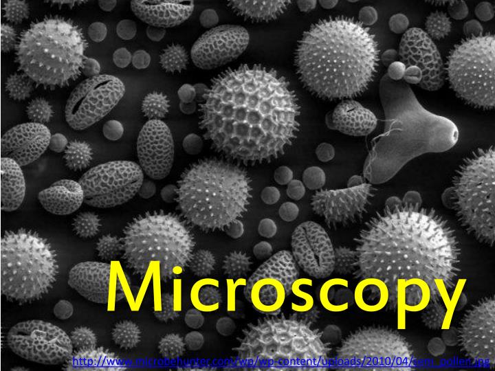
Microscopy http://www.microbehunter.com/wp/wp-content/uploads/2010/04/sem_pollen.jpg
Microscopy Microscope • the study of objects or organisms (e.g., bacteria, protists, cells, etc.) too small to be seen by the naked eye using a tool called a microscope • an instrument that gives an enlarged image of the object under study • Compound microscope • Electron microscope • Scanning probe microscope http://www.kennislink.nl/ , http://www.sciencelearn.org.nz/
Compound light microscope • Magnification • Enlargement of the image of the specimen • Total magnification = objective lens magnification * ocular lens magnification • Resolution • ability to see a gap separating two dots in an image that, to the naked eye, are not separated. • Influenced by frequency of light waves and quality of the lens • Contrast • Condenser and diaphragm modify size and intensity of a light beam http://www.cls.zju.edu.cn, http://science.howstuffworks.com
Using the microscope • Carrying the microscope properly • Mounting the slide • Viewing the specimen *images are backward and inverted *FOV gets darker as magnification increases • Preparing the microscope for storage
Preparing samples for viewing • Staining cells for better visibility • Place several drops of stain on one edge of the cover slip • The process of diffusion will allow the stain to go under the cover slip and stain the specimen • (tissue paper may be placed on the opposite edge of the cover slip to help the stain diffuse under the cover slip) http://www.biosci.ohio-state.edu, http://www.doctortee.com/dsu/tiftickjian/bio100/cell-lab.html
Microscope Math • Theoretical magnification = ocular X objective • Estimating the diameter of the field of view (LPO diam)(LPO mag) = (HPO diam)(HPO mag) • Estimating cell size = diameter / # of cells spanning the diameter http://3.bp.blogspot.com/-FfK5dESrZLM/TbwGrFcruQI/AAAAAAAAACY/F9bYyBqnqAQ/s1600/hydrilla+leaf.bmp