Download
1 / 51
540 likes | 642 Views
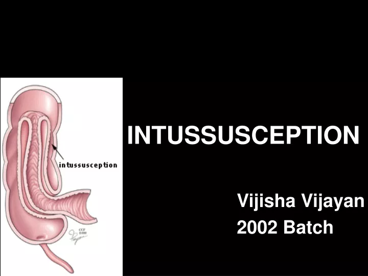
INTUSSUSCEPTION Vijisha Vijayan 2002 Batch. DEFINITION.

E N D
INTUSSUSCEPTION Vijisha Vijayan 2002 Batch
DEFINITION “INTUSSUSCEPTION IS THE TELESCOPING OF ONE PORTION OF INTESTINE INTO THE OTHER, FROM PROXIMAL TO DISTAL, BY PERISTALSIS, PULLING THE MESENTERY ALONG WITH IT.”
HISTORY • In the late 17th century, Paul Barbette described the invagination of one bowel segment into another • In 1971, Sir Jonathan Hutchison reported the first successful surgical resection
AETIOLOGY • Primary or Idiopathic – usually in children • Secondary to a pathological lead point – incidence increases with age
PATHOLOGICAL LEAD POINTS - Meckel’s diverticulum - Benign and malignant neoplasms - Appendix / appendiceal stump - Henoch-Schonlein Purpura - Jejuno-gastric Intussusception - Post-operative - Others
PATHOLOGY • Composed of three parts -Entering inner tube -Returning or Middle tube -Sheath or Outer tube *Apex - advancing part *Intussusception – mass *Neck – junction of entering layer with mass
NECK APEX
It is an example of Strangulating Obstruction as the blood supply of the inner layer is impaired • The degree of ischaemia depends on the tightness of invagination • Later may lead to Gangrene and Perforation
TYPES • Ileo colic77% • Ileo-ileo colic12% • Ileo-ileal5% • Colo colic2% • Multiple1% • Retrograde0.2%
CLINICAL FEATURES • Age5-10 months • ABDOMINAL PAIN In children, suspicion should be aroused in case of a healthy child having sudden, severe, intermittent, cramping abdominal pain. Between the episodes of pain, the child is quiet. Later the child becomes lethargic
VOMITINGBilious • RED-CURRANT JELLY STOOLS On examination, • Palpable abdominal mass, sausage-shaped, located in the right upper quadrant of abdomen, right lower region is empty – SIGN DE DANCE • Dehydration,abdominal distension • P/R Bleeding
DIAGNOSIS • History • Physical examination • Confirm by investigations
INVESTIGATIONS • Plain abdominal radiograph - air-fluid level with a paucity of gas in the right lower quadrant - sparse gas within the colon - soft tissue mass
Small bowel dilatation & paucity of gas in the right lower &upper quadrants .
Ultrasound -’Target’ of intussuscepted layers of bowel on transverse view -’Pseudo-kidney’ sign when seen longitudinally
PEUDO KIDNEY SIGN TARGET SIGN
Barium enema – both diagnostic and therapeutic Pre-requisites; -intravenous hydration -nasogastric intubation -broad-spectrum antibiotics -sedation Finding – ‘claw-sign’
Contraindications; 1). Peritonitis 2). Haemodynamic instability 3). Intussusception located fully in small intestine
TREATMENT 1) Hydrostatic Reduction Procedure - prepare patient - contrast material (Barium enema) is elevated 36 inches above the table and reduction monitored by fluoroscopy - reduction confirmed by resolution of mass, reflux of barium into proximal ileum Complication – perforation (1-3% ) Recurrence – 11%
2) Air reduction -Column of insufflated air is monitored so as not to exceed 80mmHg in infants <6 months and 120mmHg in older infants for periods of three minutes -Reduction is noted when caecal mass disappears and the small bowel becomes distended with air
3) Operative management Indications; -Failure with previous methods -Recurrence -Peritonitis -Necrotic bowel
Procedure; -Transverse muscle-cutting incision in right lower quadrant -Mass identified and manual reduction attempted by retrograde milking of the intussucepiens proximally, NEVER pull it out -Appendicectomy
REDUCTION END TO END ANASTOMOSIS
-If ischaemic and reduction not possible – LIMITED RESECTION with a primary end-to-end anastomosis -Perforation and significant fecal soiling - ENTEROSTOMY
1). MECKEL’S DIVERTICULUM • Diverticulum invaginates into intestine and is carried forward by peristalsis • May be ileoileal or ileocolic • Clinical features - urge to defaecate -early vomiting -red-currant jelly stools -palpable mass • Treatment – surgical resection
2). NEOPLASMS • Benign – common • Can cause partial or total bowel obstruction • Cramping abdominal pain • Palpable mass • Treatment - Surgery Colon: Intussusception with neoplasm - adenocarcinoma
3)APPENDIX • Rare • Difficult to diagnose – symptoms non-specific • Intussusception of appendiceal stump after appendicectomy –within 2 weeks post-operative -present as abdominal pain, vomiting, bleeding P/R, palpable mass • Diagnosis – Barium enema, CT scan • Treatment - Surgery
4). HENOCH-SCHONLEIN PURPURA • Causes sub-mucosal haematoma • Common in children
5). JEJUNO-GASTRIC INTUSSUSCEPTION • Rare, long-term complication of Billroth II surgery • Clinical features non-specific • Efferent limb, rather than afferent is the intussusceptum
Diagnosis – water-soluble upper g.i. contrast study shows ‘coiled-spring’ appearance within gastric remnant • Endoscopy will show jejunal segments as they migrate in and out of gastric remnant between episodes of intussusception • CT Scan
Treatment – Surgery 1) efferent limb can be anchored to the parietal peritoneum OR 2) a new GJ possibly using a Roux-en-Y reconstruction
6)INTERNAL INTUSSUSCEPTION • Laxity of rctal fixaton at the sacrum cause prolapse • It is a precursor to complete rectal prolapse • Incidence increased with ageing ,connective tissue disorders etc…
Symptoms • Mucous discharge • Rectal bleeding • Incomplete evacuation INVESTIGATIONS • COLONOSCOPY • BARIUM ENEMA
Treatment • Corret rectal prolapse • Glycerine suppository • Small enema SURGERY INDICATION – severe symptoms,chronic bleeding from ulcer,anal incontinance
7)POST OPERATIVE • Signs of early small bowel obstruction within a week of surgery
8)MISCELLANEOUS • Foreign body • Ectopic pancreatic or gastric tissue • Intestinal duplication
SUMMARY • Intussusception is the telescoping of one portion of intestine into the adjacent part by peristalsis pulling the mesentery along with it • Abdominal pain ,vomiting and red currant jelly stools are the classical triad • Barium enema is both diagnostic and therapeutic
Contd…. • Surgery if the above fails • The associated morbidity and occasional mortality is directly related to delay in diagnosis