Download
1 / 63
990 likes | 2.14k Views
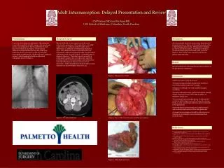
intussusception. Introduction. the telescoping or invagination of a proximal portion of intestine ( intussusceptum ) into a more distal portion ( intussuscipiens ). is one of the most common causes of bowel obstruction in infants and toddlers.

E N D
Introduction • the telescoping or invagination of a proximal portion of intestine (intussusceptum) into a more distal portion (intussuscipiens). • is one of the most common causes of bowel obstruction in infants and toddlers.
The Vascular compromise and subsequent bowel necrosis are the primary concerns with intussusceptions. • Although uncommon, in patients who undergo operative reduction of intussusception, as many as 10% may require bowel resection.
Frequency • The incidence of intussusception is 1.5-4 cases per 1000 live births. • The male-to-female ratio of 3:2. • The greatest incidence of idiopathic intussusception is in infants aged 9-24 months.
A seasonal incidence has been described, with peaks in the spring, summer, and the middle of winter. These periods correspond to peaks in the occurrence of seasonal gastroenteritis and upper respiratory tract infections.
Etiology • Intussusception is most often (80%) ileocolic , but it may be ileoileal, colocolic, or ileoileocolic. • Most infants and toddlers (95%) do not have an identifiable specific lead point.
In these idiopathic cases, careful examination may reveal hypertrophied mural lymphoid tissues (Peyer patches), which are due to adenovirus or rotavirus. • A specific lead point that draws the proximal intestine and its mesentery inward and propagates it distally through peristalsis is identified in only 5% of cases and is most commonly found in cases of ileoileal.
Specific lead points are more commonly found in children older than 3 years and almost always in adults with intussusception. • Meckeldiverticulum is the most common lead point, followed by polyps and duplications.
Other lead points described include lymphomas, submucosal hemorrhage with Henoch-Schönleinpurpura, hemangiomas, and lymphosarcomas. • Children with cystic fibrosis (CF) may present with intussusception due to inspissatedmeconium in the terminal ileum.
Presentation • The infant with intussusception has a history of severe cramping or colicky abdominal pain occurring intermittently every 5-30 minutes. • During these attacks, the infant screams and flexes at the waist, draws the legs up to the abdomen, and may appear pale.
jejunoileal or ileoileal intussusception, which usually does not have a specific lead point, accounts for approximately 1% of intussusceptions in children of all ages.
These episodes may last for only a few seconds and are separated by periods of calm normal appearance and activity. • some infants become quite lethargic and somnolent between attacks. • Early on, vomiting of undigested food may occur.
As attacks continue, emesis may turn bilious. • The Stool appears normal in early in the course of the illness then become dark red and mucoid (resembling currant jelly), a sign of intestinal ischemia and mucosal sloughing.
Disorders characterized by bowel obstruction, colicky abdominal pain, blood in the stool, an intra-abdominal mass, or a combination of these should be considered in the differential diagnosis of intussusception. • These include gastroenteritis, appendicitis, Meckeldiverticulum, malrotation with midgutvolvulus, or incarcerated hernia
Upon initial inspection, the abdomen may appear scaphoid; during paroxysms, it may be rigid; and later in the course of the illness, it may become distended with signs of peritonitis. • Careful palpation after an attack has subsided may reveal an ill-defined or sausage-shaped mass.
With early ileocolic intussusception, the mass is typically found in the right upper quadrant or abdomen. • This mass may be difficult to locate in inconsolable infants because of abdominal rigidity from muscle straining.
If episodes of cramping are witnessed, the careful examiner may auscultate peristaltic rushes in the area of the intussusception. • The rectal examination should commence with inspection of fecal material in the diaper. • Normal-appearing stool should be tested for occult blood.
The presence of mucoid or frankly bloody stool supports the diagnosis. • Rarely, inspection of the anus may reveal the prolapsed tip of the intussusception. • A digital rectal examination should be performed routinely, looking for blood or a mass higher in the anal canal.
Workup Laboratory Studies • Obtain CBC count with differential and chemistry profile. Blood chemistry abnormalities are not specific for intussusception. • The laboratory investigations may reflect dehydration, anemia, leukocytosis, or a combination of these.
Imaging Studies • Plain radiography: Early in the course of the illness, findings on plain radiographic examination of the abdomen (supine and upright) may be unremarkable. • Findings suggestive of intussusception include dilated loops of small bowel with or without air-fluid levels, an airless or opacified right lower quadrant or both.
This is an abdominal plain radiograph of a 14-week-old patient with intussusception. Note the nonspecific appearance of bowel obstruction.
Ultrasonography • First reported as a useful diagnostic tool in intussusception by Burke in 1977. • the diagnosis of intussusception has been verified by a number of authors, with a sensitivity and specificity of 100%.
Characteristic findings include a target sign visible on transverse section and a pseudo-kidney sign viewed on longitudinal section. • Sonography is best used as a diagnostic tool of exclusion when the index of suspicion for intussusception is lower.
The experience with sonography-guided hydrostatic reduction of intussusception is limited in the Western hemisphere. In Europe, successful reduction has been reported in 76-95% of cases and only one case of perforation in 825 cases.
Transverse ultrasonographic view (target sign) of intussusception
Longitudinal ultrasonographic view (pseudo-kidney sign) of intussusception
CT examination The evaluation of abdominal pain often leads to CT examination. Although not indicated for the diagnosis of intussusception, intussusception can be found incidentally on CT scan Appearance of intussusception on CT scan.
Diagnostic Procedures • Diagnostic and therapeutic enema: -It’s replacing the surgery as the initial management of stable patients. • The diagnostic enema is therapeutic in 80-90% of patients.
treatment is usually concluded in the radiology suite, and some surgeons elect to observe these patients in the hospital until they are tolerating an oral diet. • A successful therapeutic reduction must demonstrate free flow of contrast (air or barium series) proximal to the ileocecal valve.
Historically, patients in whom enema reduction was unsuccessful were taken immediately to the operating room for laparotomy and manual reduction. • in patients who are clinically stable, second and third attempts at pneumatic or hydrostatic reduction have proven effective.
author prefers to use air initially in the infant or child with suspected intussusception. • Perforation is a risk with either barium or air but poses less of a problem with air, because the combination of barium and feces may result in severe peritonitis with wide peritoneal soilage.
The technique for reduction: • In preparation for contrast study, patients should have IV access, and although not universally used, and better to have a nasogastric tube in place. • A lubricated straight catheter is placed into the rectum and secured by taping the buttocks together tightlydation may be helpful.
many radiologists prefer a balloon-tipped catheter, laceration or perforation of the rectum is a risk with balloon inflation. • A manometer and blood pressure cuff are connected to the catheter, and air is insufflated slowly to a pressure of 70-80 mm Hg (maximum 120 mm Hg) and followed fluoroscopically as it percolates proximally through the colon
The column of air stops at the intussusception, and a plain radiograph is taken. • If no intussusception exists or if the reduction is successful, air is observed to rapidly pass into the small bowel. • Another radiograph is taken at this point, and the air is allowed to escape prior to removing the catheter
At the completion of the procedure, postreduction radiography should confirm the absence of free air on supine and decubitus/upright radiographs. • Difficult reductions may require several attempts. The use of glucagon (0.5 mg/kg) for facilitating relaxation of the bowel has yielded mixed results and is not routinely used
Ultrasonography is advocated to aid in the diagnosis and assist with hydrostatic reduction of intussusception. • Studies advocating its use for diagnosis report sensitivities of 98.5-100%, specificities of 88-100%, and negative predictive values of 100%.
In 1977, a comparison of the efficacy of ultrasonographically guided versus fluoroscopically guided hydrostatic reduction in 46 patients with intussusception. • The ultrasound group had 3 recurrences (11.5%), 1 lead point (4.4%), and 19 successful reductions (73%).
Only 1 recurrence (4.2%), 1 lead point (4.4%), and 12 successful reductions (50%) occurred in the same number of patients undergoing hydrostatic reduction with barium. • No complications occurred in either group, and the accuracy rate of diagnosing a complete reduction was 100% with both forms of reduction
The authors concluded that ultrasonographically guided hydrostatic reduction for childhood ileocolic intussusception is preferred because it is safe, accurate, has a higher success rate, and can avoid radiation exposure risk.
The indication of reduction • When the barium seeps between the tow end and produces the characteristic radiological appearance of a coiled spring • The filling of the cecum is often slow and it may become quite distended for awhile before the sudden rash of the barium into the distal ileum indicative of reduction
The interval between the trial is 2to 4 will be safe(although longer interval may be triad if the Pt stable • The duration of each interval less than45 minutes
This ileocolic intussusception is observed using air-contrast enema. Intussusception has been reduced to the level of the cecum.
This ileocolic intussusception is observed using barium contrast enema. Intussusception has been reduced to the level of the proximal transverse colon.
Histologic Findings • Resected specimens show varying degrees of ischemia, necrosis, or both. • Benign reactive lymph node hyperplasia and Peyer patch hyperplasia is common. • Resected specimens should be carefully examined for potential lead points (eg, Meckeldiverticulum, polyps, lymphoma).
:Indications Stable patients with a high index of suspicion for intussusception without evidence of ischemic bowel, perforation, or sepsis may undergo immediate contrast enema for diagnosis and treatment of suspected intussusception.
Immediate surgery is indicated in unstable patients, in patients who have peritonitis, or in patients with bowel perforation during attempted enema reduction. • Elevated temperature and WBC counts have also served as relative indicators for surgery.
Patients requiring surgery must be aggressively resuscitated with fluids, and care must be taken to preserve body temperature preoperatively, intraoperatively, and postoperatively. • Contraindications to enema reduction include evidence of bowel perforation and peritonitis.
Treatment • Medical Therapy: • surgical personnel should be notified, an IV line should be inserted, and IV hydration started. • A nasogastric tube should be inserted and placed to suction.
If the patient is markedly distended or has a dilated loop of bowel, an abdominal radiograph should be obtained. • Antibiotics should be administered based on clinical suspicion of peritonitis or infection (sepsis) or in patients with a markedly elevated WBC count.