Download
1 / 4
40 likes | 70 Views
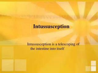
A case of 14yr old boy with an ectopic pancreatic lesion in the ileal segment presenting with intussusception is being described. Histological examination revealed ectopic pancreatic tissue in submucosa of ileum with intussusception. The unusual site and difficulty of making an accurate diagnosis is highlighted with review of literature.

E N D
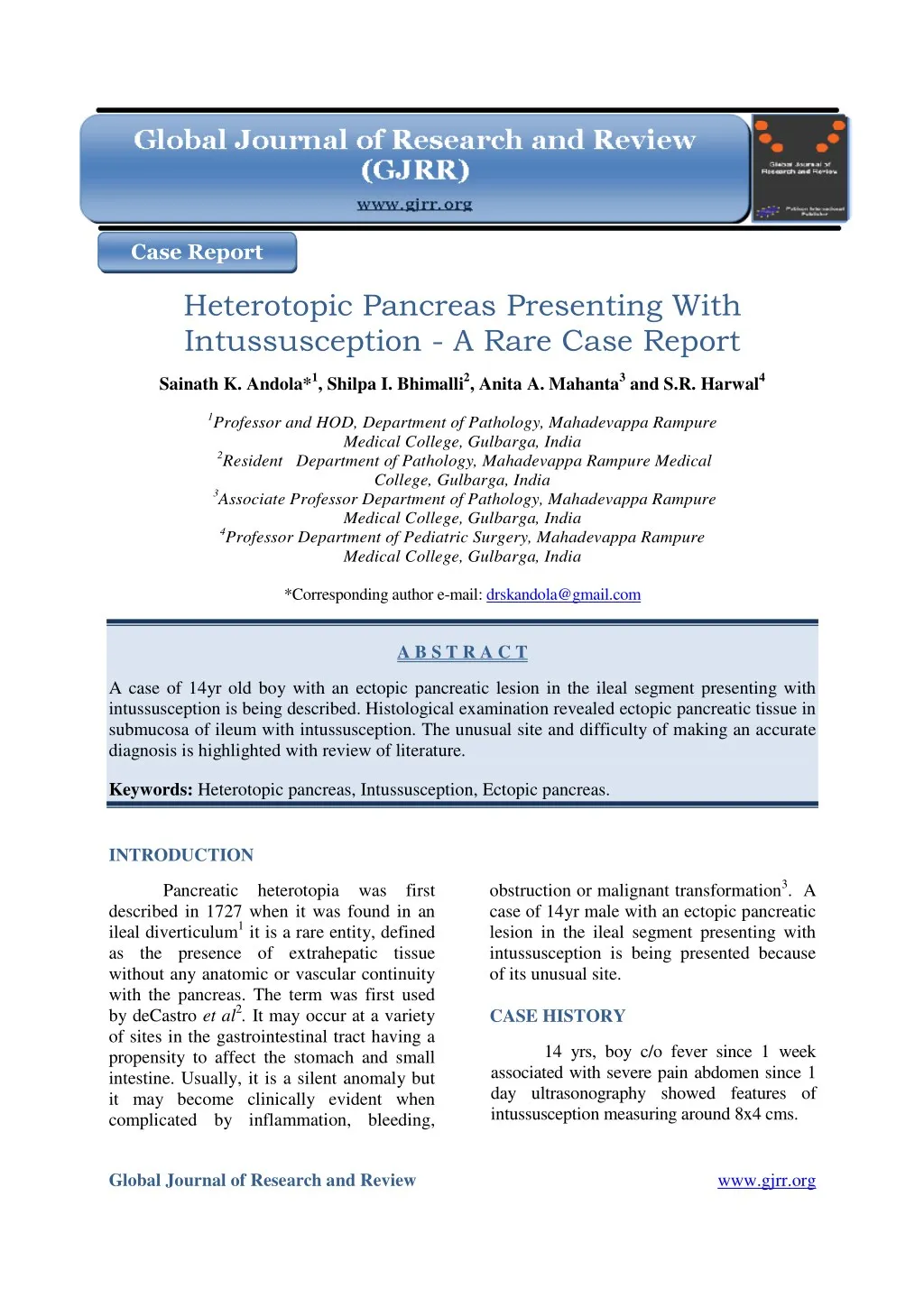
Case Report Heterotopic Pancreas Presenting With Intussusception - A Rare Case Report Sainath K. Andola*1, Shilpa I. Bhimalli2, Anita A. Mahanta3 and S.R. Harwal4 1Professor and HOD, Department of Pathology, Mahadevappa Rampure Medical College, Gulbarga, India 2Resident Department of Pathology, Mahadevappa Rampure Medical College, Gulbarga, India 3Associate Professor Department of Pathology, Mahadevappa Rampure Medical College, Gulbarga, India 4Professor Department of Pediatric Surgery, Mahadevappa Rampure Medical College, Gulbarga, India *Corresponding author e-mail: drskandola@gmail.com A B S T R A C T A case of 14yr old boy with an ectopic pancreatic lesion in the ileal segment presenting with intussusception is being described. Histological examination revealed ectopic pancreatic tissue in submucosa of ileum with intussusception. The unusual site and difficulty of making an accurate diagnosis is highlighted with review of literature. Keywords: Heterotopic pancreas, Intussusception, Ectopic pancreas. INTRODUCTION obstruction or malignant transformation3. A case of 14yr male with an ectopic pancreatic lesion in the ileal segment presenting with intussusception is being presented because of its unusual site. CASE HISTORY Pancreatic heterotopia was first described in 1727 when it was found in an ileal diverticulum1 it is a rare entity, defined as the presence of extrahepatic tissue without any anatomic or vascular continuity with the pancreas. The term was first used by deCastro et al2. It may occur at a variety of sites in the gastrointestinal tract having a propensity to affect the stomach and small intestine. Usually, it is a silent anomaly but it may become clinically evident when complicated by inflammation, bleeding, 14 yrs, boy c/o fever since 1 week associated with severe pain abdomen since 1 day ultrasonography showed features of intussusception measuring around 8x4 cms. Global Journal of Research and Review www.gjrr.org
Andola et al__________________________________________________ ISSN 2393-8854 All routine blood & urine Several theories have been proposed to explain the pathogenesis and occurrence of pancreatic heterotopia. The most tenable theory implicates that during the development of normal pancreas from several evaginations, originating from the wall of the primitive duodenum, one or more evaginations may remain in the bowel wall. Migration of this along with the development gastrointestinal tract gives rise to the ectopic pancreatic tissue9,10. Histopathologically, the heterotopic pancreatic tissue may have all the elements of the normal pancreas which include pancreatic acini, pancreatic ducts, and islets of Langerhans. Heinrich in 1909 proposed three types of heterotopic pancreas but his classification was modified by Gaspar-Fuentes in 1973 acquiring its final form. Type1 heterotopia is composed of pancreatic ducts only, referred as canalicular variety. Type 2 heterotopia is characterized by acinar tissue only (exocrine pancreas). Type 3 heterotopia is made up of islet cells only (endocrine pancreas)11. The pancreatic ectopic tissue is usually silent but can also undergo complications that occur in normal pancreatic tissue such as acute or chronic pancreatitis, abscess and pseudocyst formation. Malignant transformation may rarely occur. Pain is one of the most common symptoms. The possible explanation is that the pain is due to endocrine and exocrine function of the heterotopic pancreatic tissue, and relates to the secretion of hormones and enzymes, being responsible for inflammation or chemical irritation of the involved tissues Barium swallow study may show a typical image of a rounded filling defect with central indentation4. The lesion within the wall of the ileum may act as a lead point. This is thought to be the mechanism of intussusception. It has also been postulated that intussusception arises from local disturbance in the motility of the small intestine caused by the heterotopic examinations were done and are within normal limits. Grossly Segment of ileum received measuring 26 cms in length, central part of wall showed blackish discoloration. One of surgical margins showed a nodular lesion measuring 2x1.5cms (fig. 1). Histological Section from intestine showed large areas of haemorrhage and acute inflammatory reaction with hyperplasia of Payer’s patches. Nodular mass revealed lobular structures consisting of round to oval cells in glandular and acinar pattern separated by thin fibrous septae. The nuclei were round with bland chromatin (fig. 2). Also seen were intralobular ducts. A diagnosis of heterotopic pancreas with intussusception was made. DISCUSSION of the Isolated heterotopic pancreas in the ileum is very rare and usually asymptomatic4. It is found in about 1-2% of autopsies. Despite its congenital origin, pancreatic heterotopia was usually discovered in adults, being more common at the age of 30-50 years with a male predominance a sex ratio of 3:15. The usual location is in the stomach in 25%- 38% of the cases, duodenum in 17%-36%, jejunum in 15%-21.7% and rare in the esophagus, gallbladder, common bile duct, spleen, mesentery, mediastinum and fallopian tubes6. The incidence in pediatric age group, varies from 6 to 16 %7.In literature less than 25 cases of isolated heterotopic pancreas of ileum as leading point of intussusception in children being described. The relative frequency of a pathological lesion causing intussusception increases with age8. However, the highest frequency of detection of pathologic lesion in cases. Of intussusception is still the first year of life. The present case is described in a 14yr old boy who presented with pain abdomen due to intussusception. GJRR[1][1][2014]033-036
Andola et al__________________________________________________ ISSN 2393-8854 REFERENCES pancreas. The reported sensitivity and specificity are 87.5% respectively5. Endoscopic ultrasonography proven to be a useful adjunct in identification of pancreatic rests. CT findings are usually non specific. CT scan localize lesions with normal pancreatic distinguish ectopic pancreas from other submucosal tumors. The diagnosis may be sometimes difficult intraoperatively due to the gross similarity of pancreatic heterotopias with gastrointestinal stromal tumour (GIST), gastrointestinal autonomic nerve tumour (GANT), carcinoid, lymphoma or even gastric carcinoma. If in doubt, frozen section is very helpful to establish the diagnosis intraoperatively and to avoid unnecessary extensive operations. ultrasound showed intussusception. Exploratory laparotomy done and segment of ileum resected. CONCLUSION and 71.4%, 1.Elfving heterotopia and its clinical importance. Acta Chir Scand 1965; 130: 593-602. 2.Barbosa deCastro JJ, Dockerty MB, Waugh JM. Pancreatic heterotopia: review of the literature and report of 41 authenticated surgical cases, of which 25 were clinically significant. Surg Gynecol Obstet 1946; 82: 527.542. 3.Cullen JJ, Weydert C, Hinkhouse MM, Ritchie J, Domann FE, Spitz D, Oberley LW. The role of manganese superoxide dismutase in the growth of pancreatic adenocarcinoma. Cancer Res 2003; 63: 1297-1303. 4.Hamada Y, Yonekura Y, Tanano A, et al; Isolated Heterotopic intussusceptions; Eur J Pediatr Surg 2000;10:197-200. 5.Li –duan zheng, shao-Tao Tang, Zhi-Yong Du;Qing-Lan Ruan: heterotrophic Pancreas in a child; world journal of paediatrics; 2009; 5(2); 146-148. 6.Chandan VS, Wang heterotopia in the gastric antrum. Arch Pathol Lab Med 2004; 128: 111-12. 7.Ormarsson OT, Gudmundsdottir I, Mårvik R. Diagnosis and treatment of gastric heterotopic pancreas. World J Surg 2006; 30:1682-9. [PMID 16902740]. 8.Sarika Verma, Pankaj Bansal Vivek Manchanda, Ruchika Gupta: Isolated ileal heterotopic pancreas in a child: A clinically undetected cause intussusceptions. IJCRI. 2014; 5(2):122– 125. 9.Alic Jacobz, Akhtar Nawaz, I, Hillal Matta, Ahmed Al Salem ;Intussusception secondary to isolated Heterotopic pancreas of the ileum: a case report and review of the literature; Annals of Saudi Medicine, Vol 22, Nos 3 -4, 2002. 10.Grigorois christodoulis, dimitris zacharoulis; Heterotopic pancreas in the stomach: A case report and literature review, world journal of gastroenterology; 2007; Dec 7; 13 (45). 11.Gaspar Fuentes A, Campos Tarrech JM, Fernandez Burgui JL, Castells Tejon E, Ruiz Rossello J, Gomez Perez J, Armengol Miro G, Hastbacka J. Pancreatic has tissue but cannot pancreas causing In signs present case ileal of W. Pancreatic Isolated heterotopic pancreas in the ileum is very rare and usually asymptomatic4. The lesion within the wall of the ileum may act as a lead point. This is thought to be the mechanism of intussusception. It should always be considered in the differential diagnosis of extra mucosal gastric lesions. Despite the development diagnostic modalities, diagnosis remains challenging. Surgical excision provides symptomatic relief and is recommended if diagnostic uncertainty remains. KEY MESSAGES of modern of ileoileal Ileal heterotopic pancreas is a rare cause of intussusception at any age it is often an incidental finding and is usually not evident clinically. Hence, careful examination of the lead point of intussusceptum in resection specimens is mandatory to delineate the underlying etiology of these cases. GJRR[1][1][2014]033-036
Andola et al__________________________________________________ ISSN 2393-8854 J. Pancreatic ectopias. Rev Esp Enferm Apar Dig. 1973; 39: 255-268. Figure 1. Central part of ileum segment showing blackish discoloration Figure 2. Microscopy reveals congested mucosal layer beneath which is seen ectopic pancreatic tissue in lobules (H & E 100x) GJRR[1][1][2014]033-036