Download
1 / 31
350 likes | 980 Views
Ventilation / Ventilation Control Tests. RET 2414 Pulmonary Function Testing Module 5.0. Ventilation / Ventilation Control Tests. Objectives Calculate tidal volume and minute ventilation Describe two causes of increased ventilation Identify an abnormal V D /V T ratio.

E N D
Ventilation / Ventilation Control Tests RET 2414 Pulmonary Function Testing Module 5.0
Ventilation / Ventilation Control Tests • Objectives • Calculate tidal volume and minute ventilation • Describe two causes of increased ventilation • Identify an abnormal VD/VT ratio
Ventilation / Ventilation Control Tests • Objectives • Calculate dead space and alveolar ventilation • Describe one method for measuring breathing response to O2 • Identify the normal breathing response to carbon dioxide (CO2)
Ventilation / Ventilation Control Tests • VT, Rate, Minute Ventilation • Tidal volume (VT) is • Volume of gas inspired or expired during each respiratory cycle • Respiratory rate (f) • Number of breaths per unit of time • Minute ventilation ( ) • Total volume of gas expired per minute • alveolar ventilation ( ) • dead space ventilation ( )
Ventilation / Ventilation Control Tests • VT, Rate, Minute Ventilation VE = f x VT • Measured with volume displacement or flow-sensing spirometer .
Ventilation / Ventilation Control Tests • VT, Rate, Minute Ventilation • VT decreased in: • Severe restrictive patterns • Neuromuscular disorders • Decreased VT is usually accompanied by an increase in respiratory rate in order to maintain alveolar ventilation ( )
Ventilation / Ventilation Control Tests • VT, Rate, Minute Ventilation • Decreases in both VT and respiratory rate are often associated with respiratory center depression • Alveolar hypoventilation
Ventilation / Ventilation Control Tests • VT, Rate, Minute Ventilation • Normal respiratory rates ranges: • 10 – 20 breaths/min • Increased in: • Hypoxia • Hypercapnia • Metabolic acidosis • Decrease lung compliance • Exercise
Ventilation / Ventilation Control Tests • VT, Rate, Minute Ventilation • Normal respiratory rates ranges: • 10 – 20 breaths/min • Decreased in: • Central nervous system depression • CO2 narcosis;a condition resulting from high levels of carbon dioxide in the blood. Confusion, tremors, convulsions, and coma may occur if blood levels of carbon dioxide are too high (>70 mm Hg or higher).
Ventilation / Ventilation Control Tests • VT, Rate, Minute Ventilation • Normal minute ventilation ranges: • 5 – 10 L/min • When used in conjunction with arterial blood gases, indicates the adequacy of ventilation
Ventilation / Ventilation Control Tests • VT, Rate, Minute Ventilation • Normal minute ventilation ranges: • 5 – 10 L/min • increases in response to: • Hypoxia • Hypercapnia • Metabolic acidosis • Anxiety • Exercise
Ventilation / Ventilation Control Tests • Dead Space /Alveolar Ventilation • Dead space is the lung volume that is ventilated but not perfused by pulmonary capillary blood flow • Anatomic (conducting airways) VDan • Alveolar (non-perfused alveoli) VDA VDan + VDA = VD • VD(Respiratory or Physiologic Dead Space)
Ventilation / Ventilation Control Tests • Anatomic Dead Space
Ventilation / Ventilation Control Tests • Dead Space /Alveolar Ventilation • The portion of ventilation wasted on the conducting airways and poorly perfused alveoli is usually expressed as a ratio: VD/VT = (PaCO2 – PECO2)X 100 PaCO2 Modification of Bohr’s equation
Ventilation / Ventilation Control Tests • Dead Space /Alveolar Ventilation • For convenience VD is often estimated as equal to anatomic deadspace; VD = 1 ml/lb of ideal body weight Valid only if little or no alveolar dead space exists due to pulmonary disease
Ventilation / Ventilation Control Tests • Dead Space /Alveolar Ventilation • Normal VD/VT ratio in adults: • 0.3 or 30% (0.2–0.4 or 20%–40%) • Increases with: • Pulmonary embolism • Acute pulmonary hypertension • Decreased cardiac output • Decreases with: • Exercise (increase cardiac output and perfusion of lung apices)
Ventilation / Ventilation Control Tests • Dead Space /Alveolar Ventilation • Alveolar ventilation is the volume of gas that participates in gas exchange in the lungs per minute
Ventilation / Ventilation Control Tests • Dead Space /Alveolar Ventilation • Alveolar ventilation at rest is approximately 4 – 5 L/min • The adequacy of can only be determined with an arterial blood gas (ABG) Hypoventilation = PCO2 >45 with a pH <7.35 Hyperventilation = PCO2 <35 with a pH >7.45
Ventilation / Ventilation Control Tests • Dead Space /Alveolar Ventilation • Decreased can result from: • Increases in VD • Destruction/dilation airway walls • >FRC (air trapping/hyperinflation) • Bronchodilators • Decreases in
Ventilation / Ventilation Control Tests • Ventilatory Response to CO2 • Ventilatory response to CO2 is a measurement of the increase or decrease in caused by breathing various concentration of carbon dioxide while PaO2 is kept normal
Ventilation / Ventilation Control Tests • Ventilatory Response to CO2 • Procedure • 1-7% CO2 is breathed through either an open or closed circuit while the following are measured: • PeTCO2 • SaO2 • P100
Ventilation / Ventilation Control Tests • Ventilatory Response to CO2 • Normal response to an increased PACO2 is a linear increase in ventilation ( ) • Approximately 3 L/min/mm Hg (PCO2)
Ventilation / Ventilation Control Tests • Ventilatory Response to CO2 • Decreased in patients with: • COPD • Increased airway resistance (Raw) • Lesions in the CNS • Chemoreceptor dysfunction
Ventilation / Ventilation Control Tests • Ventilatory Response to Oxygen • Ventilatory response to O2 is a measurement of the increase or decrease in causes by breathing various concentration of O2 while PaCO2 is kept normal
Ventilation / Ventilation Control Tests • Ventilatory Response to Oxygen Procedure 20%-12% O2 is breathed through either an open or closed circuit while the following are measured: , PaO2, P100, PetCO2 The test is repeated with decreasing concentrations of O2
Ventilation / Ventilation Control Tests • Ventilatory Response to Oxygen • Normal response to a decreasing PaO2 is an exponential increase in ventilation ( ) once the PaO2 is less than 60 mm Hg (SaO2 <90%) 60 torr/90% Saturation O2
Ventilation / Ventilation Control Tests • Ventilatory Response to Oxygen • Significance and Pathology • Patients with obesity-hypoventilation syndrome, obstructive sleep apnea, and idiopathic hypoventilation will show a marked decrease response to hypoxemia
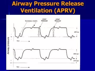
Ventilation / Ventilation Control Tests • Occlusion Pressure (P100 or P0.1) • P100 is the pressure generated during the first 100 milliseconds of inspiratory effort against an occluded airway. • It is a measurement of the neural output from the medullary centers that drive ventilation rate and volume
Ventilation / Ventilation Control Tests • Occlusion Pressure (P100 or P0.1) Normally P100 values are: • 1.5 – 5.0 cm H2O Usually measured at varying PetCO2 values or levels of O2 desaturation to assess the effect of changing stimuli to ventilation
Ventilation / Ventilation Control Tests • Occlusion Pressure (P100 or P0.1) P100 is usually plotted against PetCO2
Ventilation / Ventilation Control Tests • Occlusion Pressure (P100 or P0.1) • P100 values will normally increase with PaCO2 (hypercapnia) or PaO2 (hypoxemia) • Healthy patients typically increase occlusion pressure 0.5 to 0.6 cm H2O/mm Hg PCO2 • Patients with COPD will not increase the P100 when the PaCO2 in increased