Download
1 / 89
1.37k likes | 2.46k Views
How to Read a Head CT. (or “How I learned to stop worrying and love computed tomography”). Andrew D. Perron, MD, FACEP. EM Residency Program Director Department of Emergency Medicine Maine Medical Center Portland, ME. Andrew D. Perron, MD, FACEP. Head CT.

E N D
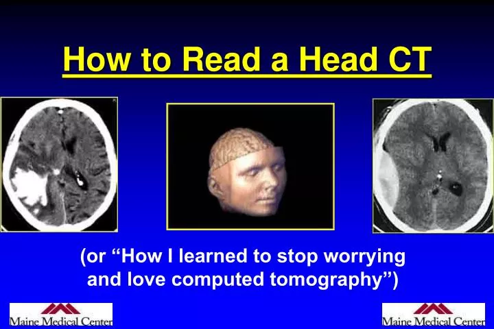
How to Read a Head CT (or “How I learned to stop worrying and love computed tomography”)
Andrew D. Perron, MD, FACEP EM Residency Program Director Department of Emergency Medicine Maine Medical Center Portland, ME Andrew D. Perron, MD, FACEP
Head CT • Has assumed a critical role in the daily practice of Emergency Medicine for evaluating intracranial emergencies. (e.g. Trauma, Stroke, SAH, ICH). • Most practitioners have limited experience with interpretation. • In many situations, the Emergency Physician must initially interpret and act on the CT without specialist assistance.
Head CT • Most EM training programs have no formalized training process to meet this need. • Many Emergency Physicians are uncomfortable interpreting CTs. • Studies have shown that EPs have a significant “miss rate” on cranial CT interpretation.
Head CT • In medical school, we are taught a systematic technique to interpret ECGs (rate, rhythm, axis, etc.) so that all aspects are reviewed, and no findings are missed.
Head CT • The intent of this session is to introduce a similar systematic method of cranial CT interpretation, based on the mnemonic…
Head CT “Blood Can Be Very Bad”
Blood Can Be Very Bad • Blood • Cisterns • Brain • Ventricles • Bone
Blood Can Be Very Bad • Blood • Cisterns • Brain • Ventricles • Bone
Blood Can Be Very Bad • Blood • Cisterns • Brain • Ventricles • Bone
Blood Can Be Very Bad • Blood • Cisterns • Brain • Ventricles • Bone
Blood Can Be Very Bad • Blood • Cisterns • Brain • Ventricles • Bone
CT Scan Basics • Introduced in 1974 by Sir Jeffrey Hounsfield. • The original “Siretom” Circa 1974
CT Scan Basics • A CT image is a computer-generated picture based on multiple x-ray exposures taken around the periphery of the subject. • X-rays are passed through the subject, and a scanning device measures the transmitted radiation. • The denser the object, the more the beam is attenuated, and hence fewer x-rays make it to the sensor.
CT Scan Basics • The denser the object, the whiter it is on CT • Bone is most dense = + 1000 Hounsfield U. • Air is the least dense = - 1000H Hounsfield U.
CT Scan Basics: Windowing Focuses the spectrum of gray-scale used on a particular image.
Posterior Fossa • Brainstem • Cerebellum • Skull Base • Clinoids • Petrosal bone • Sphenoid bone • Sella turcica • Sinuses
2nd Key Level Sagittal View 2nd Key Level
CSF Production • Produced in choroid plexus in the lateral ventricles Foramen of Monroe IIIrd Ventricle Acqueduct of Sylvius IVth Ventricle Lushka/Magendie • 0.5-1 cc/min • Adult CSF volume is approx. 150 cc’s. • Adult CSF production is approx. 500-700 cc’s per day.
CT Scans Andrew D. Perron, MD, FACEP 36
B is for Blood • 1st decision: Is blood present? • 2nd decision: If so, where is it? • 3rd decision: If so, what effect is it having?
B is for Blood • Acute blood is bright white on CT (once it clots). • Blood becomes hypodense at approximately 2 weeks. • Blood becomes isodense at approximately 1 week.
B is for Blood • Acute blood is bright white on CT (once it clots). • Blood becomes hypodense at approximately 2 weeks. • Blood becomes isodense at approximately 1 week.
B is for Blood • Acute blood is bright white on CT (once it clots). • Blood becomes hypodense at approximately 2 weeks. • Blood becomes isodense at approximately 1 week.
Epidural Hematoma • Lens shaped • Does not cross sutures • Classically described with injury to middle meningeal artery • Low mortality if treated prior to unconsciousness ( < 20%)
Subdural Hematoma • Typically falx or sickle-shaped. • Crosses sutures, but does not cross midline. • Acute subdural is a marker for severe head injury. (Mortality approaches 80%) • Chronic subdural usually slow venous bleed and well tolerated.
CT Scan Andrew D. Perron, MD, FACEP 47
CT Scan Andrew D. Perron, MD, FACEP 48
Subarachnoid Hemorrhage • Blood in the cisterns/cortical gyral surface • Aneurysms responsible for 75-80% of SAH • AVM’s responsible for 4-5% • Vasculitis accounts for small proportion (<1%) • No cause is found in 10-15% • 20% will have associated acute hydrocephalus
CT Scan Sensitivity for SAH • 98-99% at 0-12 hours • 90-95% at 24 hours • 80% at 3 days • 50% at 1 week • 30% at 2 weeks • Depends on generation of scanner and who is reading scan.