Download
1 / 90
970 likes | 1.54k Views
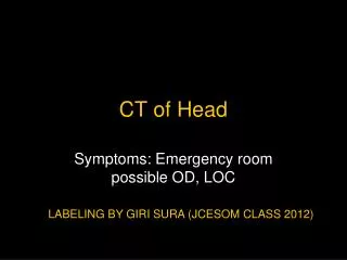
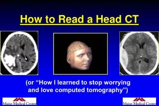
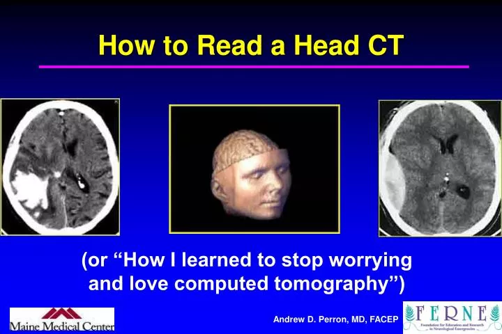
How to Read a Head CT. (or “How I learned to stop worrying and love computed tomography”). Andrew D. Perron, MD, FACEP. EM Residency Program Director Department of Emergency Medicine Maine Medical Center Portland, ME. Andrew D. Perron, MD, FACEP. 2. Head CT.

E N D
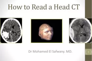
How to Read a Head CT (or “How I learned to stop worrying and love computed tomography”)
Andrew D. Perron, MD, FACEP EM Residency Program Director Department of Emergency Medicine Maine Medical Center Portland, ME Andrew D. Perron, MD, FACEP 2
Head CT • Has assumed a critical role in the daily practice of Emergency Medicine for evaluating intracranial emergencies. (e.g. Trauma, Stroke, SAH, ICH). • Most practitioners have limited experience with interpretation. • In many situations, the Emergency Physician must initially interpret and act on the CT without specialist assistance.
Head CT • Most EM training programs have no formalized training process to meet this need. • Many Emergency Physicians are uncomfortable interpreting CTs. • Studies have shown that EPs have a significant “miss rate” on cranial CT interpretation.
Head CT • In medical school, we are taught a systematic technique to interpret ECGs (rate, rhythm, axis, etc.) so that all aspects are reviewed, and no findings are missed.
Head CT • The intent of this session is to introduce a similar systematic method of cranial CT interpretation, based on the mnemonic…
Head CT “Blood Can Be Very Bad”
Blood Can Be Very Bad • Blood • Cisterns • Brain • Ventricles • Bone
Blood Can Be Very Bad • Blood • Cisterns • Brain • Ventricles • Bone
Blood Can Be Very Bad • Blood • Cisterns • Brain • Ventricles • Bone
Blood Can Be Very Bad • Blood • Cisterns • Brain • Ventricles • Bone
Blood Can Be Very Bad • Blood • Cisterns • Brain • Ventricles • Bone
CT Scan Basics • A CT image is a computer-generated picture based on multiple x-ray exposures taken around the periphery of the subject. • X-rays are passed through the subject, and a scanning device measures the transmitted radiation. • The denser the object, the more the beam is attenuated, and hence fewer x-rays make it to the sensor.
CT Scan Basics • The denser the object, the whiter it is on CT • Bone is most dense = + 1000 Hounsfield U. • Air is the least dense = - 1000H Hounsfield U.
CT Scan Basics: Windowing Focuses the spectrum of gray-scale used on a particular image.
Posterior Fossa • Brainstem • Cerebellum • Skull Base • Clinoids • Petrosal bone • Sphenoid bone • Sella turcica • Sinuses
Sagittal View C Circummesencephalic Cistern
CT Diagnostics Where is the most sensitive area to examine the CT for increased ICP? • Lateral Ventricles • IVth ventricle • Basilar Cisterns • Gyral pattern
2nd Key Level Sagittal View 2nd Key Level Circummesencephalic Cistern
CT Diagnostics Where is the most sensitive area to examine the CT for ventricular dilation? • IIIrd ventricle • IVth ventricle • Temporal horns of lateral ventricles
3rd Key Level Sagittal View Circummesencephalic Cistern
CSF Production • Produced in choroid plexus in the lateral ventricles Foramen of Monroe IIIrd Ventricle Acqueduct of Sylvius IVth Ventricle Lushka/Magendie • 0.5-1 cc/min • Adult CSF volume is approx. 150 cc’s. • Adult CSF production is approx. 500-700 cc’s per day.
CT Scans Andrew D. Perron, MD, FACEP 37
A Few Kid-Specific Thoughts • Premature Infants (30-34 weeks): Larger sylvian, basilar (circummesencephalic) cisterns. Larger subarachnoid spaces Thin cerebral cortex (Gray matter) Prominent white matter (with higher water content) Limited cortical gyral pattern Ventricles are variable: slit-like to well-developed • Term Infant (36-41 weeks): Small, slit-like lateral ventricles Continued white-matter prominence More prominent sulcal pattern Temporal horns unlikely to be seen • 1st & 2nd years of Life: Marked growth of all lobes of the brain (proportionally greatest in frontal lobes) Wide variation in lateral ventricle size (3rd and 4th fairly constant) Temporal horns unlikely to be seen.
1 day 1 year 2 years Andrew D. Perron, MD, FACEP 40
B is for Blood • 1st decision: Is blood present? • 2nd decision: If so, where is it? • 3rd decision: If so, what effect is it having?
CT Diagnostics At what point does blood become isodense with brain? • About 48 hours • About 1 week • About 2 weeks • After 1 month
B is for Blood • Acute blood is bright white on CT (once it clots). • Blood becomes hypodense at approximately 2 weeks. • Blood becomes isodense at approximately 1 week.
B is for Blood • Acute blood is bright white on CT (once it clots). • Blood becomes hypodense at approximately 2 weeks. • Blood becomes isodense at approximately 1 week.
B is for Blood • Acute blood is bright white on CT (once it clots). • Blood becomes hypodense at approximately 2 weeks. • Blood becomes isodense at approximately 1 week.
Epidural Hematoma • Lens shaped • Does not cross sutures • Classically described with injury to middle meningeal artery • Low mortality if treated prior to unconsciousness ( < 20%)
Subdural Hematoma • Typically falx or sickle-shaped. • Crosses sutures, but does not cross midline. • Acute subdural is a marker for severe head injury. (Mortality approaches 80%) • Chronic subdural usually slow venous bleed and well tolerated.
CT Scan Andrew D. Perron, MD, FACEP 50