Download
1 / 1
10 likes | 133 Views
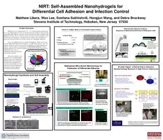
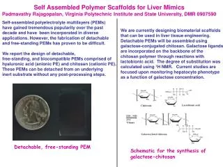
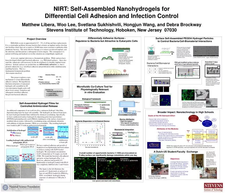
Osteoblast. (a). (f). (b). ( g ). (c). ( h ). ~1 m. (d). ~ 2 mm. ( i ). (e). ~350 m. Therapeutic Delivery/ Host defense mechanism. S. epidermidis. acidic pH. PVPON. acidic pH, after crosslinking. PMAA. a. b. (PMAA) 10 EDA S. Epidermidis 4 h.

E N D
Osteoblast (a) (f) (b) (g) (c) (h) ~1 m (d) ~2 mm (i) (e) ~350 m Therapeutic Delivery/ Host defense mechanism S. epidermidis acidic pH PVPON acidic pH, after crosslinking PMAA a b (PMAA) 10EDA S. Epidermidis 4 h (PMAA) 10 EDA + JFLO S. Epidermidis 4 h 10 μm 10 μm 100 mm 100 mm NIRT: Self-Assembled Nanohydrogels for Differential Cell Adhesion and Infection Control Matthew Libera, Woo Lee, Svetlana Sukhishvili, Hongjun Wang, and Debra Brockway Stevens Institute of Technology, Hoboken, New Jersey 07030 Differentially Adhesive Surfaces - Repulsive to Bacteria but Attractive to Eukaryotic Cells Surface Self-Assembled PEGDA Hydrogel Particles to Control Bacteria/Cell-Biomaterial Interactions Project Overview Infection occurs in approximately 0.5 – 5% of all hip and knee replacements. It is a catastrophic problem, because bacteria that colonize an implant surface develop into biofilms where they are as much as 10,000 times more resistant to antibiotics than planktonic bacteria. The most effective therapy is to remove an infected implant, cure the infection, and then pursue a subsequent revision surgery. The consequences to patient well being and medical cost in this situation are compellingly significant. At its core, implant infection is a biomaterials problem. While surfaces have been developed which repel bacterial adhesion – e.g. PEGylated surfaces – these also repel the eukaryotic cells necessary for the development of a healthy implant-tissue interface. Instead, surfaces are needed that are differentially adhesive, i.e. that it promote eukaryotic (e.g. osteoblast) adhesion and proliferation while simultaneously repelling bacteria. This is a fundamental biomaterials problem that remains unsolved. This project explores a new mechanism to create differentially adhesive surfaces. We hypothesize that heterostructures of nanosized hydrogels self assembled in 2Dover micrometer length scales willallow focal contact formation and subsequent osteoblast adhesion but prevent bacterial adhesion. PEG gel particles can deposit on the PLL modified Si wafer surface by electrical self-assembly to obtain modified surfaces with controllable gel-particle density. Lower and higher particles density surface were both tested in bacteria and osteoblast culture. Cell-Interactive nanohydrogels hierarchically structured on the surface of a macroscopically beaded surface of a modern orthopaedic implant. PLL Si UV is used to polymerize PEGDA in the DCM droplets which obtained by DCM/water emulsion. Lower PEGDA density higher PEGDA density Osteoblast 4 days culture result PEG gel-modified surface reduces short term S.epi adhesion/growth. Bacteria/Cell/Biomaterial Interactions • 4 types of substrate were studied: • bare Si; • 2 – PLL modifed Si; • Gel modified Si (low conc); • Gel modified Si (high conc); • Infection Rates • Hips 0.3 - 1% • Knees 1 - 4% • Fixation devices > 15% e.g. Intramedullary trauma rods Cytoskeleton covered substrate area is used to indicate how the cells adhere and spread. After 4 days culture, the osteoblast can still adhere and spread on the gel-modified surfaces. Microfluidic Co-Culture Tool for Physiologically Relevant in vitro Evaluation • Infection by Staphylococcal Biofilms • S. aureus (40%) • S. epidermis (20%) bare PLL high PEGDA Biological Framework Self-Assembled Hydrogel Films for Controlled Antimicrobial Release Protein Conditioning An additional component of our work involves continuous hydrogel thin films deposited using layer-by-layer self assembly. The hydrogels are derived from layer-by-layer hydrogen-bonded films stabilized by chemical crosslinking. Specifically, we have synthesized surface hydrogels by depositing poly(vinyl pyrrolidone) (PVPON)/ poly(methacrylic acid) (PMAA) multilayers at the surface of precursor-modified silicon wafers, followed by crosslinking using carbodiimide chemistry with addition of ethylene diamine ( EDA) as a crosslinker. The resulting hydrogels were loaded at pH 7.5 with an antibacterial polypeptide. Broader Impact: Nanotechnology in High Schools Implant Material Goals of the HS Outreach Effort- Expose high school students to nanotechnology-based research - Demonstrate societal relevance- Enhance and modernize topics taught in standard high school biology and chemistry • Device Attributes • High-throughput • Time-lapsed visualization • Cross contamination-free Bacteria Dispersion in 8-Channel Device Implement small pilot Develop draft modules Year 1 Attributes of the Modules- Ease of implementation in biology and chemistry courses- Minimal time requirement for implementation- Contain a hands-on or laboratory activity- Address National Science Education Standards (NSES) Biomaterial Integration Stabilization of hydrogel Implement larger pilot Revise draft modules Year 2 Finalize modules Dissemination Year 3 1 2 3 4 stabilization at basic pH Loading hydrogel with antibacterial polypeptide JFLO PMAA gel loaded w/ polypeptide JFLO We have explored adhesion and growth of Staphylococcus Epidermidis bacterial culture at surfaces coating with JLFO-loaded hydrogels. We used initial concentration 5x10⁶ colonies/mL in 3% tryptic soy broth (TSB). We found that bacterial cells adhered and grew on bare hydrogels (Fig. 1, a). However, adhesion and growth of S. Epidermidis to hydrogels loaded with JFLO was completely inhibited after 2 and 4 hours. PMAA hydrogel CIESE has nearly 20 years of K-12 curriculum and professional development expertise in STEM education, and has impacted over 20,000 educators worldwide 5 min adhesion S.epi (green) Si substrate (blue) A Dutch-US Student/Faculty Exchange PEGDA particles (black) Left: Stevens PhD student Eva Wang meeting with UMCG collaborators on flow-cell experiments. Below: Stevens undergrad Altida Patimetha working on her summer 2009 co-culture experiment at UMCG. A small number of opportunistic bacteria (1-1000) pre-inoculated on Ti alloy surface can significantly damage osteoblasts within one day. Confocal image for S.epi growing on the PEGDA modified surface. Objectives Leverage Stevens’expertise in biomaterials design, synthesis, and processing with UMCG expertise in clinically oriented physiological assessment Create international exchange opportunities for Stevens undergrads rooted in Stevens faculty research Osteoblast + 105 cfu/ml S. epidermidis Osteoblast only Osteoblast + 102 cfu/ml S. epidermidis The figure to the left illustrates the growth of S. Epidermidis at surfaces of bare (a) and JFLO-loaded (b) (PMAA)10 EDA -crosslinked hydrogels during exposure of substrates to TSB after 4 hours. Confocal images (left) and SEM (right) imaging shows good osteoblast adhesion and spreading on surfaces with cell adhesiveness modulated by PEG gel partyicles on a cell-adhesive PLL surface. cell spreading on the PEGDA modified surface. University Medical Center Groningen (UMCG) 100 mm Live (green) and dead (red) osteoblasts