Download
1 / 19
260 likes | 458 Views
Cell to Cell Junctions and Adhesion. Lecture 24 BSCI 420/421 Oct 28/29, 2002 “In our every deliberation, we must consider the impact of our decisions on the next seven generations.” - From the Great Law of the Iroquois Federation A. Cell Adhesion Molecules B. Cell-Cell Junctions

E N D
Cell to Cell Junctions and Adhesion • Lecture 24 BSCI 420/421 Oct 28/29, 2002 • “In our every deliberation, we must consider the impact of our decisions on the next seven generations.” • - From the Great Law of the Iroquois Federation • A. Cell Adhesion Molecules • B. Cell-Cell Junctions • 1. Occluding Jxs • 2. Anchoring Jxs • 3. Communicating Jxs
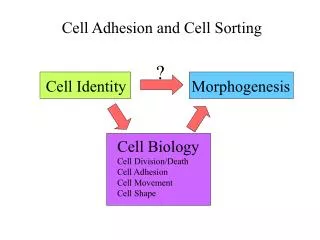
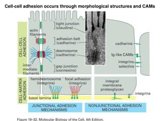
Formation of multicell organisms requires specific interaction • between cells to hold the cells together and to communicate • in order to coordinate activities. • A. 4 types of Cell Adhesion Molecules (CAMs) • are used to hold animal cells together: • Cadherins • Ig-like CAMs • Selectins • Integrins • All are single-pass transmembrane proteins anchored to the • cytoskeleton by their cytoplasmic domains.
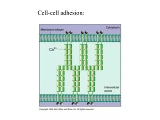
Cadherins – Ca2+ binding adhering proteins • When Ca2+ is bound to the 5 cadherin-repeat domains, • They can bind to similar domains from an adjacent cell. • Removal of extracell Ca2+ can cause dissociation of cells.
Cadherin interactions are homophilic, with identical cadherens interacting to form tissues of like cell types. Table 19-3 Classical cadherins E – epithelial & embryonic cells P – placental & epidermis N – Neural cells, lens, & muscles VE – Vascular endothelial cells Non-classical cadherins Desmocollin and desmoglein – desmosomes of epithelia like skin Protocadherins – synapses
2. Ig-like CAMs Are Ca2+ independent CAMs 20 different N-CAMs all by alternate splicing of one gene
3. Selectins Mediate transient interactions by heterophilic binding to cell surface glycoproteins on other cells.
4. Integrin Binds cells to the extracellular matrix (fibronectin or laminin) Or to Ig-family receptors. Binding can be regulated.
B. Cell to Cell Junctions are formed when groups of molecules • Interact between two cells. • 3 broad classes: • Occluding Jxs 2. Anchoring junctions 3. Communicating jxs • “ “ seal cells together to prevent passage of • material between cells. • Main type: Tight Junctions • elect-dense • tracer to • Fig 19-3 apical [ l ] or • basolateral [r]
TEM of Tight Jxs: Freeze-fracture & thin section views Fig 19-4-pt2
Recall 2 important fxs of tight jxs In glucose transport across Int. epithelia: 1. Prevent flow of Glucose between cells 2. Prevent lateral diffusion of transporters From one mem. Domain to the other
2. Anchoring Jxs a. Adherens Jxs
b. Desmosomes are circular spot junctions that produce strong adhesions between two cells. Esp. skin, intestinal epithelia, heart muscle Fig 19-11.
Diagram of desmosomes Hemidesmosomes look like ½ des, but use integrins inst. of cadherins
3. Communicating Jxs • Gap Jxs allow small molecules to flow between cells: e.g.: Ions, sugars, NTs, cAMP • 2 membranes are separated by a 2 nm gap. • Six proteins called connexins form a connexon and 2 • connexons end to end form a 1.5 nm channel between 2 cells. • Fig 19-16
The size of the gap jxn channel was determined by injecting fluorescent peptides of different sizes into Gap jx-coupled salivary gland cells of Drosophila. 1200 Da mols could pass but 2000 Da mol would not. Injection of Ca2+ along w peptides closed the channel. Why would cells want to do this?
Fxs; Fig 19-17 Synchronizing beat of heart muscle cells. Coupling oocyte to Granulosa cells Mutant Cx37 causes Infertility Oocyte needs signals from the receptors on The granulosa cells For ovulation.
Plasmadesmata serve similarly as comm. jxs in plants Fig 19-20