Download
1 / 27
270 likes | 364 Views
The Shoulder Done by : ghder ali nama. Introduction. The part of the body where the humerus attaches to the scapula. The shoulder must be mobile enough for the wide range actions of the arms and hands, but also stable enough to allow actions such as lifting, pushing and pulling.

E N D
Introduction • The part of the body where the humerus attaches to the scapula. • The shoulder must be mobile enough for the wide range actions of the arms and hands, but also stable enough to allow actions such as lifting, pushing and pulling. • It is made up of three bones. • Clavicle, • Scapula • Humerus.
Joints of the shoulder: 1. Glenohumeral joint(main one, ball and socket joint, articulation between the glenoid fossa of the scapula (shoulder blade) and the head of the humerus 2. Acromioclavicular joint(articulation between the acromion process of the scapula and the lateral end of the clavicle ) 3. Sternoclavicular joint (articulation between sternal end of the clavicle, and the manubrium sterni • There are two kinds of cartilage in the joint: 1. Articular cartilage : covers humerus head and glenoid surface. It’s a white cartilage which allows the bones to glide and move on each other. When this type of cartilage starts to wear out (a process called arthritis), the joint becomes painful and stiff. 2. Labrum : its a ring of rigid fibrous cartilage surrounding the glenoid cavity, it stabilizes the ball and socket joint!
Shoulder Movements: It is the most mobile joint in the human body. The muscles and joints of the shoulder allow it to move through a remarkable range of motion, • Armabduction • Arm adduction • Armflexion(90°) • Arm extention(45°) • Medial rotation of the arm (55°) • Lateral rotation of the arm(40_45°) • Arm circumduction (this is a combination of the above movement)
Rotator Cuff muscles • The group of four muscles and their tendons that act to stabilize the shoulder . *the strength of the joint depend on the tone of these group of muscle which across in front,above ,behind the jont 1- Supraspinatous – abducts the arm 2- Infraspinatous – external rotation 3- Teres Minor – external rotation 4- Subscapularis – internal rotation
These muscles arise from the scapula and connect to the head of the humerus, forming a cuff at the shoulder joint. They hold the head of the humerus in the small and shallow glenoid fossa of the scapula. Nerve supply:axillary and suprascapular nerve
Rotator Cuff Disorders • Tendinitis. • Tear. • Frozen Shoulder. • Instability.
Calcific Tendinitis • A disorder characterized by deposits of crytalline calcium phosphate in any tendon of the rotator cuff muscles causing inflammation and pain. • It is of unknown etiology. • Most people over the age of 40 Pain is aggravated by elevation of the arm above shoulder level or by lying on the shoulder. • sever Pain may awaken the patient from sleep. • its one of the most painful conditions in the shoulder .
When this condition is symptomatic, it may present in the following 2 ways: • Chronic, relatively mild pain with intermittent flares, similar to shoulder impingement syndrome, is believed to indicate that the condition is in the formative phase. • Mechanical symptoms may arise from a large calcific deposit → build up of pressure in the tendon→intense pain→limitation of movement
Diagnoses by : • Xray • Ultrasound (more accurate) calcific deposits are visible as lumps or cloudy areas. Mostly found on the greater tuberosity
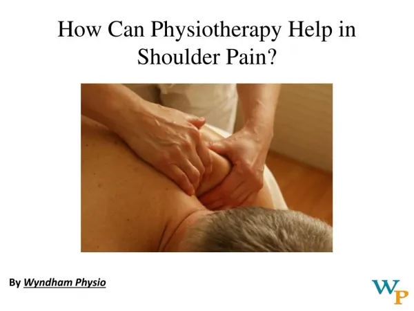
Treatment: • NSAID injection • Injections, needling, and lavage Breaking up the calcific deposits by repeatedly puncturing them with a needle, aspirating the calcific material, with the help of saline. • Surgery(rarely required) • Physiotherapy to regain muscle strength
Impingement Syndrome (chronic tendinitis) • Also called: swimmer's shoulder or thrower's shoulder • It is a clinical syndrome which occurs when the tendons of the rotator cuff muscles become irritated and inflamed as they pass through the subacromial space, the passage beneath the acromion. • Individuals at highest risk are laborers and those working in jobs that require repetitive overhead activity like swimmers and athletes. • Symptoms: pain increase at night, weakness and loss of movement at the shoulder • increase in shoulder pain with overhead activities
Causes: The rotator cuff muscle tendons pass through a narrow space between the acromion process of the scapula and the head of the humerus. Anything which causes further narrowing of this space can result in impingement syndrome. This can be caused by bony structures such as subacromial spurs (bony projections from the acromion), osteoarthritic spurs on the acromioclavicular joint, and variations in the shape of the acromion. Thickening or calcification of the coracoacromial ligament can also cause impingement. Loss of function of the rotator cuff muscles, due to injury or loss of strength, may cause the humerus to move superiorly, resulting in impingement. Inflammation and subsequent thickening of the subacromial bursa.
Treatment: Conservative mostly : • injectable corticosteroid • Ice packs • Cessation of painful activity and rest • physiotherapy . • If the patient remains significantly disabled and has no improvement after 3 months of conservative treatment, • consider other etiologies or refer for surgical evaluation.
Rotator Cuff tears • Rotator cuff tears are tears of one or more of the four tendons of the rotator cuff muscles . • Rotator cuff tears are among the most common conditions affecting the shoulder • The most frequent cause of rotator cuff damage is age related degeneration and less frequently by sports injuries or trauma • the supraspinatus muscle is most frequently torn as it passes below the acromion; • the tear usually occurs at its point of insertion onto the humeral head at the greater tuberosity
Clincal Features • Age:45-75 year old. • Acute tears: raising arm against resistance, (like in weight lifting,) or falling forcefully, causes Immediate pain that radiates through the arm, and limited range of motion, specifically during abduction motions of the shoulder . • Drop Arm sign. (The result is positive if the patient is unable to lower the affected arm slowly and smoothly from a position of 90 degrees of abduction. The arm drops immediately to the side).
Types: Partial tear: recover gradually With supraspinatus tendonitis • Complete tear: • Sudden shoulder strainor a complication of tendonitis • pain soon subside • grossweakness of abductor muscles
Treatment: • Conservative • NSAID injections • Rest • Physiotherapy • Operative - young active individuals with complete tears. - contraindicated in elderly
Mri for rotator cuff tears www.icareunit.com
Thank you www.icareunit.com