Download
1 / 50
540 likes | 1.03k Views
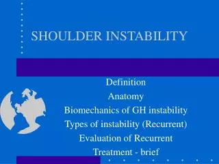
Pediatric Shoulder Instability. Mark M. Scheffer, M.D. Orthopaedic Surgeon Dartmouth-Hitchcock Concord. Shoulder Anatomy. General Anatomy:. Shoulder Anatomy. Glenohumeral Ligaments:. Shoulder Anatomy. Glenohumeral Ligaments. Shoulder Anatomy. Coracoacromial Arch:. Shoulder Stability.

E N D
Pediatric Shoulder Instability Mark M. Scheffer, M.D. Orthopaedic Surgeon Dartmouth-Hitchcock Concord
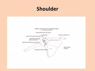
Shoulder Anatomy • General Anatomy:
Shoulder Anatomy • Glenohumeral Ligaments:
Shoulder Anatomy • Glenohumeral Ligaments
Shoulder Anatomy • Coracoacromial Arch:
Shoulder Stability Definition: Static and Dynamic Forces that Interact to Keep the Humeral Head Centered in the Glenoid in order to Maintain Shoulder Function.
Shoulder Stability • Static and Dynamic Stabilizers
Shoulder Stability • Static Stabilizers: • Bone – glenoid and humeral head • Coracoacromial Arch • Labrum • Capsuloligamentous Complex
Shoulder Stability • Dynamic Stabilizers • Rotator Cuff • Deltoid, Trapezius, Lat Dorsi
Shoulder Instability • Classification: Traumatic vs. Atraumatic
Traumatic vs. Atraumatic • TUBS vs. AMBRI • TUBS: • Traumatic • Unidirectional • Bankart Tear • Surgical Treatment C. Parker, U Tenn
Traumatic vs. Atraumatic • TUBS vs. AMBRI • AMBRI • Atraumatic • Multidirectional • Bilateral • Rehab • Inferior Capsular Shift
Traumatic vs. Atraumatic Unidirectional vs. Multidirectional
Traumatic Dislocation • Bankart Lesion:
Traumatic Dislocation • Bankart Lesion:
Traumatic Dislocation • Bankart Lesion:
Traumatic Dislocation • Boney Bankart Lesion:
Traumatic Dislocation • Hill-Sachs Lesion:
Traumatic Dislocation • Hill-Sachs Lesion:
Traumatic Dislocation • Associated Injuries • Rotator Cuff Tear • Axillary Nerve Injury • Fracture – tuberosities, Salter, Glenoid
Atraumatic Dislocation: • Capsule is Large, Stretched Out • Other Shoulder is also Loose • Ankle Sprains, Other Dislocations
Traumatic vs. Atraumatic • Evaluation: • History • Physical Exam • Imaging • Treatment
History • When did first dislocation occur? • How Many? • What Causes the Dislocation? • PT? • Other Joint Problems?
History • Voluntary Component?
Physical Exam • Generalized Laxity • Examine both shoulders • Range of Motion • Neuro Exam – Axillary Nerve! • Special Tests
Physical Exam • Sulcus Sign:
Physical Exam • Apprehension Sign:
Physical Exam: • Relocation Test:
Physical Exam • Impingement Test : (Kids Don’t Get Primary Impingement!)
Xrays: • AP IR and ER:
Xrays • Scapular Y and Axillary
Xrays • West Point Axillary View:
Xrays Dislocation:
Xrays • Boney Bankart:
Xrays: • Watch Out:
MRI • Always Get an MRI/Arthrogram – Gadolineum!
MRI • Multidirectional Instability:
MRI • Bankart Tear:
Treatment - Traumatic • Acute Reduction:
Treatment - Traumatic • Sling 1 – 3 Weeks, Rehab • Very high rate of recurrence in Teens (up to 90%) • Case can be made for primary Bankart Repair
Treatment - Traumatic • Surgical: • Arthroscopic Bankart • Open Bankart • Possible w/ Capsular Imbrication
Treatment - Traumatic • Surgical:
Treatment - Atraumatic • Rehab: • Rotator Cuff Strengthening • Scapular Stabilization Exercises TIME! – Tends to get better as people age
Treatment - Atraumatic • Surgical – • Arthroscopic Capsular Imbrication • Capsular Shift (arthroscopic or open)
Recurrent Dislocations • Bone Involvement: • Humerus • Increased Hill Sachs Lesion size • Glenoid • Anteroinferior Bone/Cartilage Loss
Recurrent Dislocations • Bone Loss - glenoid
Recurrent Dislocations • Bone Loss – Glenoid and Humeral Head:
Posterior Dislocations • Seizures – can be missed (21/24 Zagreb 2003) • Electricution • Muscle Imbalance
Posterior Dislocations • Reverse Hill-Sachs:
Shoulder Instability Thanks!