Download
1 / 43
490 likes | 863 Views
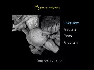
SURGICAL MANAGEMENT OF BRAINSTEM GLIOMA. Brainstem tumor classification systems. Brainstem tumor classification systems. Choux el al., 2000 CT & MRI Type I: diffuse Tape II: intrinsic, focal Type Ill: exophytic , focal Type IV: cervicomedulIary. DIFFUSE BSG.

E N D
Brainstem tumorclassification systems Choux el al., 2000 CT & MRI Type I: diffuse Tape II: intrinsic, focal Type Ill: exophytic, focal Type IV: cervicomedulIary
DIFFUSE BSG • Diffuse BSG- • Hypo on T1, hyper on T2, inhomogenous contrast enhancement within or around the tumour. • Contrast enhancement in only 1/3rd cases. • No significant difference in prognosis with/without contrast enhancement.
FOCAL BSG • Focal BSG • Well circumscribed, of limited size, may be partially cystic • Midbrain >Medulla>Pons • Hypo on T1, hyper on T2, nidus of focal enhancement • Usually pilocyticastrocytomas
DORSAL EXOPHYTIC BSG • Dorsally exophytic BSG- • Intra-IV th ventricular tm • Resemble vermian astrocytoma with involvement of IV th ventricular floor
Intraoperative monitoring • Cranial nerves- • EMG monitoring – III,IV,V,VI,VII, IX,X,XI,XII • BAEP – VIII nerve • SSEP and MEP - tracts
SSEP – Monitors Dorsal column integrity Stimulation sites UL – Median/Ulnar nerves LL -- Posterior tibial nerve >= 50% decrease in amplitude and associated 10% increase in latency – significant
BAEP Representation of electrical activity generated by the eighth cranial nerve and brainstem in response to auditory stimulation.
BAEP Wave V is the most reliably seen wave A shift in latency of 1 millisecond or a drop in amplitude of 50% could be significant. changes occur, may be due to improper retraction on the cerebellum and brain stem. Reversible with change of position of the retractors
Choice of approach • Location of lesion • Area to which lesion come close to pial surface • Clinical status of patient • Comfort of individual surgeon
Midbrain /Thalamus Pontomesencephalic 1.OZ 2. Lateral SCIT Pons Lateral SCIT 1. CPA. Retrosigmoid2. Medial MCP - suboccipital ± telovelar3. Lateral/high MCP - Retrosigmoid/lateral SCIT Pontomedullary Cervicomeduullary 1. Retrosigmoid2. Far lateral 1. Suboccipital2. Far lateral
Planned approach for ventral lesions Ventral / lateral rostral to cranial nerve V Transsylvian / subtemporal Between lower nerve and cranial Presigmoid / retrosigmoid nerve V Caudal to lower group Far lateral CHEN ET AL- NEUROSURGERY VOLUME 68 I NUMBER 31 MARCH 2011,
Planned approach for dorsal lesions Dorsal Midbrain Suboccipitaltranstentorial/ supracerebellarinfratentorialFloor of fourth ventricle Transcerebellomedullary fissure Medulla intertonsillar CHEN ET AL - NEUROSURGERY VOLUME 681 NUMBER 31 MARCH 2011,
Safe entry zones to brainstem - Rationale The 2-point method was used as an objective means to choose the surgical approach One point is placed in the center of the lesion, and a second point is placed either where the lesion comes closest to a pial surface or at the safest entry point into the brainstem.
PONS • Ventral and ventro-lateral pons approached by • Retro Sigmoid approach • Pre sigmoid approcah • Trans petrosal approach
Safe entry zone for ventro lateral pons RecaldeR. MICROSURGICAL ANATOMY OF THE SAFE ENTRY ZONES ON THE ANTEROLATERAL BRAINSTEM RELATED TO SURGICAL APPROACHES TO CAVERNOUS MALFORMATIONS. Neurosurgery, 2008. • Peritrigeminal zone between emergence of fifth and seventh nerve located medially to fifth and lateral to pyramidal tract. • Fibersare directed horizontally so myelotomy should be in horizontal direction. • Surgical window – • Horizontal – 4.64mm ( 3.8 – 5.6 mm) • Vertical - 11.2 mm (9.5- 13.1mm)
Suprafacial Triangle • 1 cm longitudinal incision from edge cerebellar peduncle and 5mm lateral to median sulcus • Length of incision –7mm MLP 1cm Cerebellar peduncle Kmn Facial nerve KyoshimaK,Kobayashi S et al.A study of safe entry zones via the floor of the fourth ventricle for brain-stem lesions. Report of three cases. JNS 1993
Infrafacial Triangle 1 cm longitudinal incision above striaemedullaris and 5 mm lateral to median sulcus Facial colliculus Abducens PFFF Facial nerve 1cm MLF Facial 4-5 mm Striaemedullares KyoshimaK,Kobayashi S et al.A study of safe entry zones via the floor of the fourth ventricle for brain-stem lesions. Report of three cases. JNS 1993
Ventral medulla • Safe corridors – • at level of retro olivarysulcus • between cranial nerve 12 and C1 at level of anterolateral sulcus • Surgical window – • cranio-caudal - 13.5 mm • Transverse - 7 mm • antero-dorsal - 2.5mm
Dorsal medulla • Safe corridors – • Posterior median fissure - below obex ,between nucleus of gracile fasciculus • posterior intermediate sulcus – between gracile and cuneatefascile • Posterior lateral sulcus - between cuneatefascile medially and spinal trigeminal tract and nucleus laterally
Ventral Midbrain • Safe entry zone – lateral to cranial nerve III , between SCA and PCA and medial to pyramidal tract Bricolo A, TurazziS:Adv Tech Stand Neurosurg 22:261– 341, 1995
Ventro lateral midbrain • Safe entry zone is lateral mesencephalic sulcus (LMS) • Minimum working distance – 4.9 mm • Maximum working distance – 11.7 mm • Mean +- SD - 8.2 +- 1.76 mm
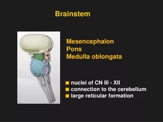
Surgical technique- • Almost all BSTs are dorsally located, therefore should be approached through posterior fossa. • Position-prone(preferred)/sitting • Midline skin incision • Suboccipitalcraniotomy±cervicallaminotomy • Y –shaped duralopening
Surgical technique- • Vermiscoagulated and split at appropriate level. • Cerebellum held to the sides using self- retaining retractors ( avoid excessive side retraction – pseudobulbarpalsy ). • IVth ventricle approached after division of medullary velum.
Tumour decompression • Conventionalsuction technique frequentlycauses brainstem dysfunction manifested by bradycardia & arrhythmia. • CUSA causes movement of adjacent structures only within 1mm of vibrating tip, allowing extensive and quick dissection adjacent to or within the substance of brainstem.
Surgical technique- Focal tumour • Rostral & caudal pole of the tumor be completely exposed. • Incision at an area where tumor is most superficial. • It also must be away from the midline and at least 1.5cm rostral to the obex - avoids injury to cr. nv nuclei X-XII. • Incision < 1cm.
Surgical technique- Focal tumor Use of plated bayonet(very small plates at the tip) as ‘microretractor’. CUSA at a low setting. Careful identification of white matter interface. Minimal manipulation of adjacent normal tissue.
Surgical technique- Cervicomedullarytumor • Suboccipital craniotomy + osteoplastic laminotomy. • Expose both rostral and caudal extent of the tumor. • USG guidance to know extent of tumor prior to opening the dura - entire tumor should be within the confines of the operative exposure.
Surgical technique- Cervicomedullarytumour • Midline myelotomy • ‘True’ midline to be identified • Identify DREZ bilaterally • If tumor is solid-cystic, myelotomyto be placed first at tumor–cyst junction and cyst is removed prior to tumor excision. • If tumor is non-cystic, myelotomywhere tumor is most voluminous & closest to the pial surface.
Surgical technique- Cervicomedullarytumour Myelotomy to be terminated 1 cm proximal to the caudal pole of the tumor → tumor is least voluminous here, removed by gradual upward dissection. At the rostral pole, tumorinvariably subpial and bulging posteriorly at the obex.
Surgical technique- Cervicomedullarytumour • USG to guide the extent of tumor excision- to confirm bulk of tumoris removed. • Don’t chase small questionable fragments. • If deterioration of SSEP/MEP during the procedure, interrupt the dissection and move to another area.
Surgical technique-Cystic tumour • Bulge into the IV ventricle. • “Collapse” of the cyst cavity and surrounding neural tissue following cyst evacuation → difficulty in identifying the solid nodule. • ‘Hand-held’ retractor compared to fixed. • Avoid frequent manipulation of retractor.
Surgical technique- Dorsally exophytictumor • Major technical complication-injury to neural structures immediately below the ependymal lining. • Remove tumor “flush” with the floor of IV ventricle • Do not pursue tumor inside the brainstem. • Facial colliculus injury
Peri & post-operative care • Perioperative steroids( methylprednisolone) • Elective ventilation for at least 48 hours • Mechanical ventilation till recovery of ventilation & normal cough reflex • LCN paresis- NG/feeding gastrostomy • V,VII nv paresis- temporary tarsorrhaphy • Good nursing care • Physiotherapy • Post-op brainstem injury mostly reversible if surgical technique is proper
Role of stereotactic biopsy • Majority of focal, dorsally exophyticand cervicomedullaryBSG are benign and resectableby direct surgery with low morbidity and good outcome.
Role of stereotactic biopsy • Reserved to • When the diagnosis is uncertain, to rule out inflammatory pathology like TB. • Focal intrinsic endophytic lesion- well limited masses within the brainstem surrounded by neural tissue and therefore do not reach the surface.
Journal quotes • ”An aggressive surgical approach to patients with intrinsic brainstemtumors significantly improves overall survival(OS) and progression free survival (PFS) compared to adult non-surgical series.” • (OS) rate is 61% and progression free survival (PFS) is 45% at 5 years. Neurosurgery: August 2012 doi: 10.1227/01.neu.0000417806.84351.f0
Take home message • Knowledge of safe entry zones. • Diffuse tumor almost invariably malignant and should not be operated upon→ Direct RT + CT • Focal lesions : If laterally located & appears to be approachable with acceptable risks, resection is appropriate. • Radical excision possible • If more centrally located → Stereotactic biopsy + Irradiation