Download
1 / 91
920 likes | 1.18k Views
Liver, Pancreas and Gallbladder. Your tutor: Rachel Boggus Boggusrl@email.uc.edu. Review. What is an exocrine gland? What is an endocrine gland?. Review. What is an exocrine gland? Retains connection to original epithelial layer, secretes product into ducts that lead to lumen.

E N D
Liver, Pancreas and Gallbladder Your tutor: Rachel Boggus Boggusrl@email.uc.edu
Review • What is an exocrine gland? • What is an endocrine gland?
Review • What is an exocrine gland? • Retains connection to original epithelial layer, secretes product into ducts that lead to lumen. • What is an endocrine gland? • Lose connection with epithelial layer and secrete products into surrounding CT and mesenchyme
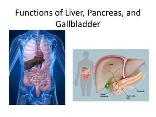
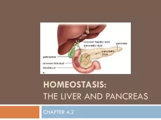
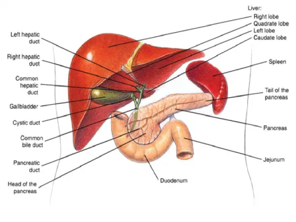
Cont. • The liver, gallbladder, and pancreas are like big glands that sprouted from the digestive tract.
Circulation to and from Liver • Where does blood get to the liver from (2 spots)? • What is a portal system? • Why have a hepatic portal system?
Circulation to and from Liver • Where does blood get to the liver from? • From the hepatic artery (20%--O2 rich) • From the hepatic portal vein (80%--O2 poor, nutrient rich) • What is a portal system? • Systemic circulation includes two capillary beds • Why have a hepatic portal system? • Because the liver is important for many functions including detox and packaging of the nutrients that we eat.
Liver blood supply cont. • Where do the blood vessels reach the liver? • Where do the portal vein and hepatic artery empty? • How does blood leave the liver?
Liver blood supply cont. • Where do the blood vessels reach the liver? • Inferior side of liver at the porta hepatis • Where do the portal vein and hepatic artery empty? • Branch through CT and empty into the sinusoids (O2 Rich and poor blood MIX here!) • How does blood leave the liver? • Tributaries that drain into hepatic veins inferior vena cava.
The Liver Lobule • What is in the corners of the lobules? • What is contained in this structure?
The Liver Lobule • What is in the corners of the lobules? • Portal triad • What is contained in this structure? • Branch of hepatic artery, branch of hepatic vein, and a bile duct (also lymph vessels and autonomic nerves)
Liver Lobule Cont. • Liver lobules consist of plates of hepatocytes • Where are the plates directed? • What are the spaces between the hepatocytes called? • Trace the venous drainage from the liver
Liver Lobule Cont. • Liver lobules consist of plates of hepatocytes • Where are the plates directed? • Toward a central vein • What are the spaces between the hepatocytes called? • Bile canaliculi—they are continuous with bile ducts in portal triad. • Trace the venous drainage from the liver • Hepatic vessels (artery and portal vein) liver sinusoidscentral veinsublobular veinshepatic veins
Liver Sinusoids • Are discontinuous—what does that mean? • What is on the surface of hepatocytes close to sinusoids? • What is the Space of Disse? • What are Kupffer cells? Where are they found?
Liver Sinusoids • Are discontinuous—what does that mean? • Composed of incomplete endothelial lining with an incomplete basal lamina • What is on the surface of hepatocytes close to sinusoids? • Lots of microvilli • What is the Space of Disse? • Space created between endothelial cells of sinusoids and microvilli of hepatocytes. • What are Kupffer cells? Where are they found? • Macrophages of the liver, found on luminal surface of endothelial cells of liver sinusoids
Bile Canaliculi • Walls formed by hepatocytes themselves • Hepatocytes have microvilli that project into lumen of canaliculi • Bile flows to the outer portions of the lobules to the bile duct in the portal triad • Bile tract lined by cuboidal cells w/in CT surrounding lobulebile ductules—join at right angles with bile ducts within portal triad • What is the epithelium of the bile ducts?
Bile Canaliculi • Terminal tubes of biliary tree • Walls formed by hepatocytes themselves • Hepatocytes have microvilli that project into lumen of canaliculi • Bile flows to the outer portions of the lobules • Bile tract lined by cuboidal cells w/in CT surrounding lobulebile ductules—join at right angles with bile ducts within portal triad • What is the epithelium of the bile ducts? • Cuboidal/columnar surrounded by CT sheath
Hepatocytes • What organelles do they contain?
Hepatocytes • What organelles do they contain? • Mitochondriaeosinophilia • RERbasophilia • SER • Peroxisomes • Golgi • Free ribosomes • Glycogen • Lipid droplets
What are the 9 functions of the hepatocyte • Protein synthesis—albumin, prothrombin, fibrinogen, lipoproteins (products released into space of Disse) (ENDOCRINE) • Bile Secretion—EXOCRINE product, secreted into biliary tree • Blood Filtration—Old RBCs broken down by Kupffer cells • Excretion—bilirubin (product of Hb breakdown) • Metabolic Storage—carbs glycogen • Metabolic functions—lipids and AAsglucose
9 fncs of hepatocytes cont. • Detoxification and inactivation—nitrogenous wastesurea (SER) • Synthesis of VLDL—main carrier of lipids in the body • Recycling IgA—from sinusoids to bile canaliculi (transcytosis)secretory IgA
CT Stroma • W/in lobules very sparse, consisting of reticular fibers (chicken wire or tree branch appearance) • Collagen borders lobules
Classic lobules, portal lobules and liver acini • What is the difference between the three?
Classic lobules, portal lobules and liver acini • What is the difference between the three? • Classic lobules—central structure central vein, portal triads at corners • Portal lobule—triangle with portal triad in center and central vein at each corner—used when thinking of exocrine (biliary) function of the liver • Liver acinus—oval shaped with central veins at apices (blood flow model)
Why do we care about the crap on the previous slide? • Because it helps you realize what hepatocytes are exposed to what things. • I.E. hepatocytes closer to the wall of the classic lobule will get more levels of O2 and nutrients and hepatocytes closer to central vein will see more of the endocrine products
Regeneration • Your liver is cool because if you take out 75% of it, it will grow back • BUT, if you are an alcoholic you get too much CT regeneration which is called cirrhosisfewer hepatocytes and poor liver structure. Too much CT, too few functional cells
Gallbladder • What are gallstones? Why are they bad?
Gallbladder • What are gallstones? Why are they bad? • Gallstones are precipitations of cholesterol, calcium carbonate, and bilirubin. Bad cause they can block bile flowjaundice and pain.
Gallbladder mucosa • Innermost layer—simple columnar epithelium—cells responsible for? How? • Lamina propria—what is it like? Why?
Gallbladder mucosa • Innermost layer—simple columnar epithelium—cells responsible for? How? • Concentration of bile by active transportof Na+ ions across basolateral memb. and into interstitial tissue. H20 follows by osmosis • Lamina propria—what is it like? Why? • Highly vascular to allow for H20 reabsorption
Gallbladder muscular and outer layers • Muscular layer—thin and irregular • What causes contraction of muscular layer? • Outer layer—serosa or adventitia?
Gallbladder muscular and outer layers • Muscular layer—thin and irregular • What causes contraction of muscular layer? • CCK • Outer layer—serosa or adventitia? • This is a TRICK, its BOTH • adventitia when buried against the liver • serosa on side facing viscera
Pancreas • Also mixed exocrine/endocrine function like liver Exocrine pancreas: • What are the secretory cells of the pancreas? • What do they secrete?
Pancreas • Also mixed exocrine/endocrine function • Exocrine pancreas • What are the secretory cells of the pancreas? • Acinar cells • What do they secrete? • Digestive enzyme precursors
Acinar cells • What do they look like? • What causes them to release their product?
Acinar cells • What do they look like? • Typical protein secreting cells • Round nucleus in lower ½, prominent nucleolus, basophilic basal cytoplasm b/c of tons of RER, large supranuclear golgi which make zymogen granules near apical surface. • What causes them to release their product? • CCK
Pancreas Ducts • Beginnings intercalated ducts composed of centroacinar cells • What do these cells do? • Trace the flow of products from the intercaleted ducts to the intestine
Pancreas Ducts • Beginnings intercalated ducts composed of centroacinar cells • What do these cells do? • Respond to secretin and release bicarb to buffer acidic chyme • Trace the flow of products from the intercaleted ducts to the intestine • Intercalatedintralobularinterlobular (in septa)
Endocrine Pancreas • Islets of Langerhans • Very pale b/c they contain less RER, contain fenestrated capillaries • What are the three types of secretory cells?
Endocrine Pancreas • Islets of Langerhans • Very pale b/c they contain less RER, contain fenestrated capillaries • What are the three types of secretory cells? • Alpha—secrete glucagon • Beta—secrete insulin • Delta—secrete somatostatin