Download
1 / 15
220 likes | 402 Views
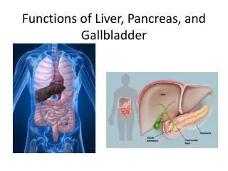
Pancreas, Liver, and gallbladder. Metallic 0 Mind. Pancreas. connective tissue forms septa. Which subdivide the gland into lobule. Exocrine secretion:. Duct System. Produce proenzymes. 40 to 50 acinar cells form acinus. Acinar cells: Shaped like truncated pyramid.

E N D
Pancreas, Liver, and gallbladder Metallic 0 Mind
Pancreas • connective tissue forms septa. Which subdivide the gland into lobule.
Exocrine secretion: Duct System Produce proenzymes. • 40 to 50 acinar cells form acinus. • Acinar cells: • Shaped like truncated pyramid. • Lie on the basal lamina. • Basal, rounded nucleus. • Basophilic cytoplasm. • Apex has secretory granules (acidophilic) • Basal cell membrane have receptors for CCK and acetylcholine. • Abundance of RER, Mictochondria, polysomes. • Centroacinar cells: • In the lumin of acinus. • Low cuboidal. • Have receptors for secretinand acetylcholine. • No myoepithelial cells.
Endocrine Pancreas • Islets of langerhans : • Surrounded by reticular fibers. • Greater number in the tail region of the pancreas
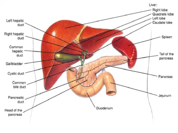
Classic lobules • Connective tissue elements (portal tracts) arrange hepatocytes in hexagon-shaped lobules (classical lobules). Classical lobules The place where 3 classical lobules are in contact is called portal area (triads)
Portal Area Parenchyma of the liver hepatocytes Hepatic artery Hepatic artery Inlet arteriole Limiting plate (modified hepatocytes) separate portal area from the parenchyma of the liver Space of Möll separate limiting plate from the conncective tissue of portal area Distributing arteriole hepatocytes
Pathway for central vein Cells are radiating from central vein forming plates of cells separated by sinusoids • Interlobular bile duct are vascularized by peribiliary capillary plexus • Central vein: • At the central of lobule. • Tributary of hepatic veins.
Hepatic sinusoids Spaces between hepatocytes Have two types of cells • Kupffer Cells: • Associated with the sinusoidal lining cells. • Phagocytic cells. • Have filopodia-like pojections • Sinusoidal lining cells: • Leaving gap between them. • The cells themselves have fenestrae. No basement membrane
Hepatic sinusoids Hepatocytes • Hepatic Stellate cells: • Known as Ito cells and fat storing cells. • Functions: • Store vitamin A • Manufacture and release type III collagen. • Secrete growth factor. • Form fibrous connective tissue Narraow space between them known as perisinusoidal space of Disse Basal lamina is absent
Hepatic Ducts Pathway for bile in liver • Composed of : • Hepatocytes • Low cuboidal cells • Occasional oval cells • Composed of : • Low cuboidal cells • Some ovoid cells
Hepatocytes • Polygonal cells • acidophilic Plasma membrane have two domains Other hepatocytes hepatocyte sinusoids • 1-Lateral domain: • Respnsible for formation of bile canaliculi. • Leakage of bile is prevented by tight junction (fasciae occludentes). • Hepatocytemicrovilli project into bile canaliculi. • Hepatocytesplasmalemmais the wall forbile canaliculi. • Have isolated gap junction to communicate with other cells. • 2- Sinusoidal Domain: • Have microvilli.
Gallbladder Mucosa is highly folded into ridges IF Not invested: adventitia Loose connective tissue Simple columnar epithelium Obliquely oriented No goblet cells no muscularis mucosa Invested by peritoneum: serosa