Download
1 / 30
300 likes | 310 Views
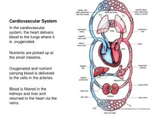
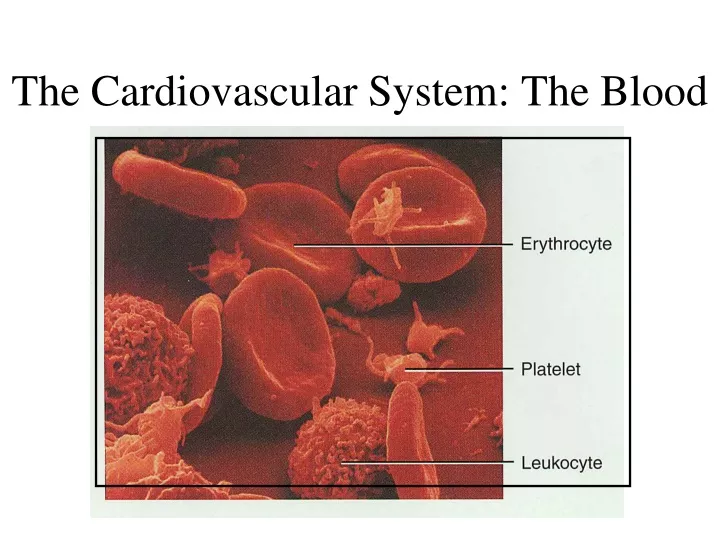
The Cardiovascular System: The Blood. Fluids of the Body. Cells of the body are serviced by 2 fluids blood composed of plasma and a variety of cells transports nutrients and wastes interstitial fluid bathes the cells of the body

E N D
Fluids of the Body • Cells of the body are serviced by 2 fluids • blood • composed of plasma and a variety of cells • transports nutrients and wastes • interstitial fluid • bathes the cells of the body • Nutrients and oxygen diffuse from the blood into the interstitial fluid & then into the cells • Wastes move in the reverse direction
Functions of Blood • Transportation • O2, CO2, metabolic wastes, nutrients, heat & hormones • Regulation • helps regulate pH through buffers • helps regulate body temperature • Protection from disease & loss of blood
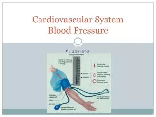
Physical Characteristics of Blood • Thicker (more viscous) than water and flows more slowly than water • Temperature of 100.4 degrees F • pH 7.4 (7.35-7.45) • 8 % of total body weight • Blood volume • 5 to 6 liters in average male • 4 to 5 liters in average female
Components of Blood • Whole blood • 55% plasma • 45% cells • 99% RBCs (hematocrit) • < 1% WBCs and platelets
Blood Plasma • 0ver 90% water • 7% plasma proteins • 2% other substances
Formed Elements of Blood • Red blood cells (erythrocytes) • White blood cells (leukocytes) • granular leukocytes • neutrophils • eosinophils • basophils • agranular leukocytes • lymphocytes = T cells, B cells, and natural killer cells • monocytes • Platelets (special cell fragments)
Formation of Blood Cells • Most blood cell types need to be continually replaced • die within hours, days or weeks • process of blood cells formation is hematopoiesis • In adult • occurs only in red marrow of flat bones like sternum, ribs, skull & pelvis and ends of long bones
Stages of Blood Cell Formation • Pluripotent stem cells • .1% of red marrow cells • replenish themselves as they differentiate into either myeloid or lymphoid stem cells • Myeloid stem cell line of development continues: • progenitor cells(colony-forming units) no longer can divide and are specialized to form specific cell types • example: CFU-E develops eventually into only red blood cells • next generation is blast cells • have recognizable histological characteristics • develop within several divisions into mature cell types • Lymphoid stem cell line of development • pre-B cells & prothymocytes finish their develop into B & T lymphocytes in the lymphatic tissue after leaving the red marrow
Hemopoietic Growth Factors • Regulate differentiation & proliferation • Erythropoietin (EPO) • produced by the kidneys increase RBC precursors • Thrombopoietin (TPO) • hormone from liver stimulates platelet formation
Red Blood Cells or Erythrocytes • Contain oxygen-carrying protein hemoglobin that gives blood its red color • 1/3 of cell’s weight is hemoglobin • Biconcave disk • increased surface area/volume ratio • flexible shape for narrow passages • no nucleus or other organelles • no cell division or mitochondrial ATP formation
Hemoglobin • Globin protein consisting of 4 polypeptide chains • One heme pigment attached to each polypeptide chain • each heme contains an iron ion (Fe+2) that can combine reversibly with one oxygen molecule
Transport of O2 and CO2 • Each hemoglobin molecule can carry 4 oxygen molecules from lungs to tissue cells • Hemoglobin transports CO2 waste from tissue cells to lungs for release • One of the breakdown products of hemoglobin is bilirubin (yellow-orange pigment). • Bilirubin enters the bloodstream and is transported to the liver where it is secreted by hepatocytes into bile.
RBC Life Cycle • RBCs live only 120 days • wear out from bending to fit through capillaries • no repair possible due to lack of organelles • Worn out cells removed by fixed macrophages in spleen & liver • Breakdown products are recycled
Erythropoiesis: Production of RBCs • Proerythroblast starts to produce hemoglobin • Many steps later, nucleus is ejected & a reticulocyte is formed • orange in color with traces of visible rough ER • Reticulocytes escape from bone marrow into the blood • In 1-2 days, they eject the remaining organelles to become a mature RBC
WBC Anatomy and Types • All WBCs (leukocytes) have a nucleus and no hemoglobin • Granular or agranular classification based on presence of cytoplasmic granules made visible by staining • granulocytes are neutrophils, eosinophils or basophils • agranulocytes are monocyets or lymphocytes
Neutrophils (Granulocyte) • Nuclei = 2 to 5 lobes connected by thin strands • older cells have more lobes • young cells called band cells because of horseshoe shaped nucleus (band) • Majority of circulating WBCs • Direct actions against bacteria • release lysozymes which destroy/digest bacteria
Eosinophils (Granulocyte) • Nucleus with 2 or 3 lobes connected by a thin strand • Slows down inflammation caused by basophils
Basophils (Granulocyte) • Irregular, s-shaped, bilobed nuclei • Involved in inflammatory and allergy reactions
Lymphocyte (Agranulocyte) • Dark, oval to round nucleus • B cells • destroy bacteria and their toxins • produces antibodies • T cells • attack viruses, fungi, transplanted organs, cancer cells & some bacteria • Natural killer cells • attack many different microbes & some tumor cells • destroy foreign invaders by direct attack
Monocyte (Agranulocyte) • Nucleus is kidney or horse-shoe shaped • Largest WBC in circulating blood • does not remain in blood long before migrating to the tissues • differentiate into macrophages • fixed group found in specific tissues • alveolar macrophages in lungs • kupffer cells in liver • wandering group gathers at sites of infection • Destroy microbes and clean up dead tissue following an infection
WBC Physiology • Less numerous than RBCs • 1 WBC for every 700 RBC • Only 2% of total WBC population is in circulating blood at any given time • rest is in lymphatic fluid, skin, lungs, lymph nodes & spleen
Platelet (Thrombocyte) Anatomy • Disc-shaped, 2 - 4 micron cell fragment with no nucleus • Platelets form in bone marrow from megakaryocytes • Short life span (5 to 9 days in bloodstream) • Help heal damaged blood vessels
Blood Clotting • Blood drawn from the body thickens into a gel • gel separates into liquid (serum) and a clot of insoluble fibers (fibrin) in which the cells are trapped • If clotting occurs in an unbroken vessel is called a thrombosis • Substances required for clotting are Ca+2, enzymes synthesized by liver cells and substances released by platelets or damaged tissues • Clotting is a cascade of reactions resulting in the formation of fibrin threads
Overview of the Clotting Cascade • Prothrombinase is formed after damage occurs • Prothrmobinase converts Prothrombin to thrombin • Thrombin converts fibrinogen into fibrin threads • Fibrin pulls damaged blood vessel together
Intravascular Clotting • Thrombosis • clot (thrombus) forming in an unbroken blood vessel • forms on rough inner lining of BV • if blood flows too slowly (stasis) allowing clotting factors to build up locally & cause coagulation • may dissolve spontaneously or dislodge & travel • Embolus • clot, air bubble or fat from broken bone in the blood • pulmonary embolus is found in lungs • can cause a stroke
Blood Groups and Blood Types • RBC surfaces are marked by genetically determined glycoproteins & glycolipids • Universal Donor? • Universal Recipient?
ABO Blood Groups • Based on 2 glycolipid antigens called A and B found on the surface of RBCs • display only antigen A -- blood type A • display only antigen B -- blood type B • display both antigens A & B -- blood type AB • display neither antigen -- blood type O