Download
1 / 24
240 likes | 654 Views
2. The Spinal Cord- A highway traveled by sensory nerve impulses headed for the brain, and by motor nerve impulses destined for the spinal nerves.Anatomy of the Spinal Cord-Meninges- connective tissue coverings that surround brain and spinal cord. Spinal meningesCranail meningesDura Mater- Most superficial meninges layer. A sac runs from Foramen Magnum to S2. Continuous with meninges of the brain. Epidural Space- located between dura

E N D
1. 1
CHAPTER 13
THE SPINAL CORD & SPINAL NERVES
KENNETH E. MULLER, PhD,PT
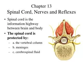
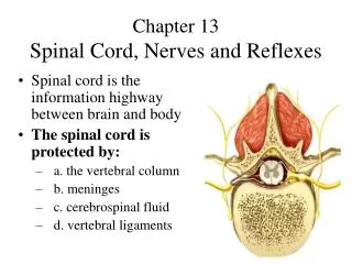
2. 2 The Spinal Cord- A highway traveled by sensory nerve impulses headed for the brain, and by motor nerve impulses destined for the spinal nerves.
Anatomy of the Spinal Cord-
Meninges- connective tissue coverings that surround brain and spinal cord.
Spinal meninges
Cranail meninges
Dura Mater- Most superficial meninges layer. A sac runs from Foramen Magnum to S2. Continuous with meninges of the brain.
Epidural Space- located between dura & vertebral canal.
3. 3 Arachnoid- middle layer. Similar to a spiders web, collagen fibers & elastic fibers. Continuous with the arachnoid of the brain. Between the dura and arachnoid is the subdural space which contains interstitial fluid.
Pia mater- Deepest meninges layer. Thin transparent layer of connective tissue that adheres to the surface of the spinal cord & brain. Full of blood vessels.
Subarachnoid space- located between arachnoid and pia. Contains CSF.
Meningitis- Inflamation of the meninges.
Deniculate ligaments- thickenings in the Pia. Protect the spinal cord from shock & sudden displacement.
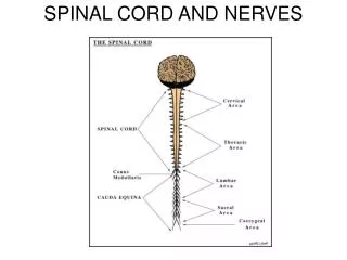
4. 4 Spinal Cord- Extends from the medulla to ~ L2
Cervical enlargement- at ~ C4-T1. Contains UE nerves.
Lumbar enlargement- at ~ T9-T12. Nerves to the lower limbs.
Conus Medularis- at end of spinal cord at ~ L2.
Filum Terminale- Extension of pia mater that anchors spinal cord to the sacrum.
Cauda Equina- roots after the spinal cord ends.
Spinal nerves- 2 nerves from each spinal segment. Named based on their location. 8 cervical, 12 thoracic, 5 lumbar, 5 sacral, 1 coccyx.
5. 5 Spinal nerves- Paths of communication between spinal cord and parts of the body.
2 bundles of axons, roots, connect nerve to a segment of the cord.
Posterior, Dorsal root- Sensory only. Conducts impulse from periphery to CNS. Has a swelling, dorsal root ganglion, which contains the cell bodies.
Anterior, Ventral root- Motor. Contains axons which conduct impulse from CNS to organs & cells.
Spinal tap- Withdraw CSF for diagnostic purposes or to introduce an anesthetic. Done between L3 & 4. Iliac crests.
6. 6 Internal Spinal Cord Anatomy-
Anterior median fissure
Posterior median sulcus
Gray mater- cell bodies, unmyelinated axons, dendrites etc. Neuroglia.
Central canal- continuous with 4th ventricle, CSF
Anterior gray horn- cell bodies of motor cells
Posterior gray horn- sensory nuclei
Anterior, ventral, white columns & Posterior, dorsal, white columns- contain bundles of nerve axons called tracts.
Sensory (ascending) tracts conduct impulses toward the brain.
Motor (descending) tracts away from brain.
7. 7 Sensory & Motor Tracts-
Sensory information travels 2 main routes-
2 spinothalamic tracts (anterior & lateral)- pain, temperature, deep pressure, touch.
Posterior columns- Proprioception, 2 point discrimination, vibration, pressure, discriminative touch.
Motor tracts, 2 types of descending pathways, direct & indirect.
Direct- Corticospinal (lateral & anterior) & corticobulbar. Convey nerve impulses destined to voluntary skeletal muscles.
Indirect- Impulses for automatic movements, postural coordinate movements, maintain tone, equilibrium.
8. 8 3 main levels of neural integration in the somatosensory system.
Receptor level
Sensory receptors transform a stimuli into and electrical response. Transduction
Generate a receptor potential, a local graded potential
Can and will experience adaptation. Pain receptors do not adapt well
Circuit level
Ascending pathways conduct sensory impulses upward through chains of 3 successive neurons.
First order neurons- cell bodies reside in a dorsal root or cranial ganglion. Synapse with second order neurons.
9. 9 Second order neurons
Cell bodies reside in the dorsal horn or medulla.
Transmit impulses to the Thalamus or Cerebellum where they synapse.
Third order neurons
Located in the Thalamus.
Conduct impulses to the somatosensory cortex of the cerebrum
Main Ascending pathways
Small diameter pain fibers synapse in the Substantia Genatinosa in the dorsal horn.
Somatosensory information is conveyed along 3 pathways
Nonspecific Ascending Pathway
Specific Ascending Pathway
Spinocebellar Tract
10. 10 Nonspecific pathway
Receive input from many different receptors and make multiple synapses in the brain stem
Also called Anterolateral pathways
Largely formed by Lateral and anterior spinothalamic pathways.
Most of the fibers transmit pain, temperature and coarse touch.
Crossover of these fibers occurs in the spinal cord.
Heavily involved in emotional aspects of perception.
Specific pathway
Also called the lemniscal system
Formed by paired tracts of the dorsal white columns
Fasciculus Gracilis and Cuneatus
Mediate precise straight through transmission
From the Thalamus impulses are forwarded to the somatosensory cortex.
Perceptual level
Neuronal circuits in the cerebral cortex
11. 11 Spinocerebellar tracts
Anterior and Posterior
Convey information from proprioceptors to the cerebellum which uses this information to coordinate skeletal muscle activity.
No conscious sensation
Perceptual level
Neuronal circuits in the cerebral cortex projected from the Thalamus
Sensory input typically evokes a behavioral response.
Perception detection- Simplest level of perception.
Detecting a stimulus has occurred
Magnitude Estimation
The ability to detect how much of the stimulus is acting on the body.
12. 12 Spatial Discrimination
Allows us to identify the site or pattern of the stimulation
2 point discrimination test
Feature Abstraction
The mechanism is which a neuron or circuit is tuned to one feature in preference to another.
Allows us to identify texture or shape
Quality discrimination
The ability to discriminate the individual qualities of an object
Pattern Recognition
The ability to take in the scene around us and recognize a familiar pattern.
13. 13 Motor pathways-
Direct motor pathways- Pyramidal pathways, nerve impulses for voluntary contraction.
Upper motor neurons- medial to the anterior horn cell. 90% of fibers decussate at the medulla. The other 10% eventually cross over at lower levels.
Lower motor neurons- anterior horn cell and out. Also called final common pathway because they are the only neurons that carry info from CNS to skeletal muscle fibers.
Lateral corticospinal tract- skilled movements of the limbs, hands and feet.
14. 14 Anterior corticospinal tract- neck, trunk and coordinating trunk movements.
Corticobulbar tract- Cranial nerves movement of eyes, tongue, neck, chewing, facial expression and speech.
Flaccid paralysis vs spastic paralysis.
Extrapyramidal tracts- all motor tracts other than anterior and lateral corticospinal.
15. 15 Spinal nerves- 31 pairs each named from where they originate.
Each nerve has 2 connections to the cord, an anterior and posterior root. Meet to form a nerve at the intervertebral foramen
Covering of a spinal nerve-
Endoneurium- covers individual axons.
Bundles of endoneurium called fascicles.
Fascicles are wrapped in perineurium.
Superficial covering over entire nerve is epineurium.
Branches- After exiting through the foramen a spinal nerve splits into branches called rami.
Posterior Rami- Dorsal side, deep muscles.
Anterior Rami- Upper & lower limbs, ventral side.
16. 16 TIME TO REST YOUR BRAIN CELLS
17. 17 Plexuses- A network of nerve fibers. (Telephone Station)
Intercostal nerves- T2-T12
Cervical Plexus- C1- C4 7 some help from C5
Supplies skin & muscles of the head & neck as well as superior shdrs, chest & diaphragm.
Phrenic nerve injury- C3,4,5
Ansa Cervicalis
18. 18 Brachial Plexus- C5-T1. R,T,D,C,B.
Axillary nerve- Deltoid
Musculocutaneous nerve- Elbow flexors
Radial nerve- Posterior arm & forearm
Median nerve- Anterior arm & some hand.
Ulnar nerve- medial arm, hand intrinsics.
Injuries-
Erb�s (brachial plexus) palsy
Radial nerve injury or entrapment
Median nerve injury or entrapment
Ulnar nerve injury or entrapment
long thoracic nerve injury
The brachial plexus will be an essay on the next test. See pg. 433.
19. 19 Lumbar plexus- L1-L5.
Sacral plexus- L5-S4
Lumbar plexus injuries-
Femoral nerve injury- anterior thigh
Obturator nerve injury- medial thigh
Sciatic nerve- Actually 2 nerves tibial & common peroneal bound together by connective tissue. Splits at the knee into tibial & common peroneal. Injuries cause foot drop with inversion.
20. 20 Dermatomes- An area of skin that provides sensory input via the posterior root of a spinal nerve. Like a road map. Knowing the dermatome helps the clinician to find the affected area
Spinal cord injuries- Most are incomplete
Quadriplegia
Hemiplegia
Paraplegia
21. 21 Neuritis- irritation of a nerve.
Shingles- Herpes zoster after chicken pox. Dermatomal in nature. Virus affects posterior root ganglion. Characterized by severe dermatomal pain, blisters, skin discoloration.
Poliomyelitis- Affects anterior horn cells. Post polio syndrome- Progressive weakness of muscles, slow degeneration. Triggered by prolonged bed rest, a fall, a minor accident or surgery. Physical therapy helps.
22. 22 Reflexes- fast, predictable, automatic responses to a change in environment.
Reflex arc- path followed by impulse to produce a reflex.
Stretch reflex-
Flexor reflex-
Crossed extensor reflex-
Plantar flexion reflex-
Babinski reflex
23. 23 Proprioceptive sensations-
what degree muscles are contracted, joint position, head orientation.
Movement of 1 body part in relation to another.
Kinesthesia- The perception of body movements.
Proprioceptors- the receptors for proprioception. Adapt slowly & only slightly.
Muscle spindles- specialized groups of muscle fibers interspersed and oriented parallel to skeletal muscle fibers.
Spindles are anchored to the endomysium and perimysium.
Consists of Intrafusal muscle fibers- contract when stimulated by medium diameter A fibers called Gamma motor neurons.
24. 24 Surrounding the muscle spindle are regular skeletal muscle fibers called extrafusal fibers innervated by large A fibers. Cell bodies located in the anterior gray horn cell.
Adjust degree and response to stretch.
Monitor change in muscle length. Information arrives at cortex allowing perception of limb position then passes to cerebellum to aid in coordination.
Tendon organs- proprioceptors found at the junction of tendon & muscle.
When tension is applied at the tendon impulses are generated to the CNS. Provide info about change in muscle tension.
25. 25 THAT�S IT
NEXT IS THE BRAIN