Download
1 / 23
320 likes | 466 Views
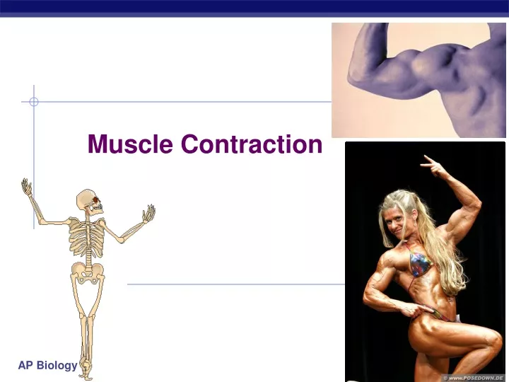
Muscle Contraction. Organization of Skeletal muscle. skeletal muscle. plasma membrane. nuclei. tendon. muscle fiber ( cell ). myofibrils. myofilaments. Structure of striated skeletal muscle. Muscle Fiber muscle cell divided into sections = sarcomeres Sarcomere

E N D
Organization of Skeletal muscle skeletal muscle plasma membrane nuclei tendon muscle fiber (cell) myofibrils myofilaments
Structure of striated skeletal muscle • Muscle Fiber • muscle cell • divided into sections = sarcomeres • Sarcomere • functional unit of muscle contraction • alternating bands of thin (actin) & thick (myosin) protein filaments
Muscle filaments & Sarcomere • Interacting proteins • thin filaments • braided strands • actin • tropomyosin • troponin • thick filaments • myosin
Thin filaments: actin • Complex of proteins • braid of actin molecules & tropomyosinfibers • tropomyosin fibers secured with troponin molecules
Thick filaments: myosin • Single protein • myosin molecule • long protein with globular head bundle of myosin proteins: globular heads aligned
Thick & thin filaments • Myosin tails aligned together & heads pointed away from center of sarcomere
sarcomere sarcomere Interaction of thick & thin filaments • Cross bridges • connections formed between myosin heads (thick filaments) & actin (thin filaments) • cause the muscle to shorten (contract)
formcrossbridge releasecrossbridge shortensarcomere Where is ATP needed? binding site CleavingATP ADP allows myosin head to bind to actin filament thin filament(actin) myosin head ADP thick filament(myosin) 1 2 ATP So that’s where those10,000,000 ATPs go! Well, not all of it! 1 1 3 1 1 4
Closer look at muscle cell Sarcoplasmicreticulum Transverse tubules(T-tubules) Mitochondrion multi-nucleated
Ca2+ ATPase of SR Muscle cell organelles • Sarcoplasm • muscle cell cytoplasm • contains many mitochondria • Sarcoplasmic reticulum (SR) • organelle similar to ER • network of tubes • stores Ca2+ • Ca2+ released from SR through channels • Ca2+ restored to SR by Ca2+ pumps • pump Ca2+ from cytosol • pumps use ATP There’sthe restof theATPs! But whatdoes theCa2+ do? ATP
Muscle at rest • Interacting proteins • at rest, troponin molecules hold tropomyosin fibers so that they cover the myosin-binding sites on actin • troponin has Ca2+ binding sites
The Trigger: motor neurons • Motor neuron triggers muscle contraction • release acetylcholine (Ach) neurotransmitter
Nerve trigger of muscle action • Nerve signal travels down T-tubule • stimulates sarcoplasmic reticulum (SR) of muscle cell to release stored Ca2+ • flooding muscle fibers with Ca2+
Ca2+ triggers muscle action • At rest, tropomyosin blocks myosin-binding sites on actin • secured by troponin • Ca2+ binds to troponin • shape changecauses movement of troponin • releasing tropomyosin • exposes myosin-binding sites on actin
How Ca2+ controls muscle • Sliding filament model • exposed actin binds to myosin • fibers slide past each other • ratchet system • shorten muscle cell • muscle contraction • muscle doesn’t relax until Ca2+ is pumped back into SR • requires ATP ATP ATP
Put it all together… 1 2 3 ATP 7 4 6 ATP 5
How it all works… • Action potential causes Ca2+ release from SR • Ca2+ binds to troponin • Troponin moves tropomyosin uncovering myosin binding site on actin • Myosin binds actin • uses ATP to "ratchet" each time • releases, "unratchets" & binds to next actin • Myosin pulls actin chain along • Sarcomere shortens • Z discs move closer together • Whole fiber shortens contraction! • Ca2+ pumps restore Ca2+ to SR relaxation! • pumps use ATP ATP ATP
Fast twitch & slow twitch muscles • Slow twitch muscle fibers • contract slowly, but keep going for a long time • more mitochondria for aerobic respiration • less SR Ca2+ remains in cytosol longer • long distance runner • “dark” meat = more blood vessels • Fast twitch muscle fibers • contract quickly, but get tired rapidly • store more glycogen for anaerobic respiration • sprinter • “white” meat
Muscle limits • Muscle fatigue • lack of sugar • lack of ATP to restore Ca2+ gradient • low O2 • lactic acid drops pH which interferes with protein function • synaptic fatigue • loss of acetylcholine • Muscle cramps • build up of lactic acid • ATP depletion • ion imbalance • massage or stretching increases circulation
Diseases of Muscle tissue • ALS • amyotrophic lateral sclerosis • Lou Gehrig’s disease • motor neurons degenerate • Myasthenia gravis • auto-immune • antibodies to acetylcholine receptors Stephen Hawking
Botox • Bacteria Clostridiumbotulinum toxin • blocks release of acetylcholine • botulism can be fatal muscle
Rigor mortis • So why are dead people “stiffs”? • no life, no breathing • no breathing, no O2 • no O2, no aerobic respiration • no aerobic respiration, no ATP • no ATP, no Ca2+ pumps • Ca2+ stays in muscle cytoplasm • muscle fibers continually contract • tetany or rigor mortis • eventually tissues breakdown& relax • measure of time of death