Download
1 / 12
160 likes | 377 Views
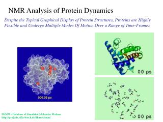
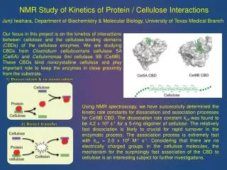
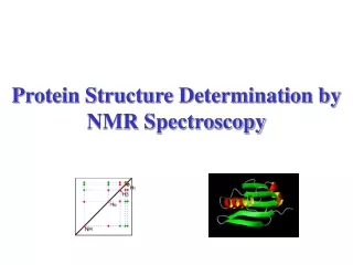
Protein NMR. Nuclear Magnetic Resonance. NMR is an analytical technique in which magnetic nuclei absorb energy from an applied electromagnetic pulse and radiate the energy back NMR is used for identifying functional groups and imaging. Quantum theory.

E N D
Nuclear Magnetic Resonance • NMR is an analytical technique in which magnetic nuclei absorb energy from an applied electromagnetic pulse and radiate the energy back • NMR is used for identifying functional groups and imaging
Quantum theory • Subatomic particles “spin” along their axis. • Atoms with paired spins have no net overall spin. • Atoms can have a net overall spin if the number of protons or number of protons and neutrons is odd. • These atoms have multiple orientations • Examples: 13C, 15N
In the absence of an external magnetic field, these orientations have the same energy. • In the presence of an external magnetic field, the energy level splits • The lower energy level contains more nuclei that the higher energy level.
Lower energy nuclei can be excited into the higher energy level by electromagnetic radition. • The frequency of the radition corresponds to the energy difference between the two nuclei states (transition frequency). • After absorption, nuclei relax to lower energy state
Chemical Shift • When the magnetic field at the nucleis is not equal to the applied magnetic field, the nucleus is sheilded by surrounding electrons. • Sheilding: If electrons produce a field opposing the applied magnetic field, the applied field strength must increase in order to reach the transition frueqncy • This results in upfield shift • Desheilding: If the electrons produce a field with the applied magnetic field, the applied field strength must decrease in order to reach the transition frequency. • This results in downfield shift
Protein Structure • Primary structure is a sequence of amino acids linked by peptide bonds • Each amino acid has a unique side chain (R); there are 20 total • Peptide bonds formed by dehydration synthesis (highlighted in red) • Chemical interactions of R groups dictate higher structure
Protein Structure Secondary structure is the formation of alpha helicies and beta sheets Tertiary structure is the 3D folding of the protein Quaternary structure is the assembly of multiple protein subunits Myoglobin from E.Coli (PDB ID: 1CQ2)
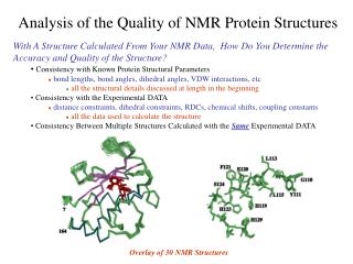
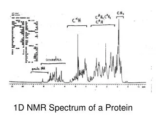
Protein NMR • Series of 2D NMR techniques • First is usually 15N-HSQC which results one signal per amino acid residue • Additional peaks from amino acids containing nitrogen in their side chains Diagram of Interactions study by other NMR techniques including HNCO (b), HNCA (c), HBHA(CBCACO)NH (d) and CBCANH (e)
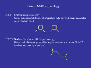
Heteronuclear Single Quantum Coherence (HSQC) • Hydrogen nuclei are excited and the energy is transferred to a neighboring 15N • The chemical shift is evolved on the nitrogen • Energy is transferred back to the hydrogen for detection. • Mainly show H-N correlations • Amino acids with NH bonds in side chains also have additional peaks Transfer of Energy between an excited hydrogen nuclei to a neighboring 15N.
Advantages & Disadvantages • Advantages • “Fingerprint”: each protein has a unique pattern • Identification of possible problems due to multiple conformation of sampel heterogenity • Disadvantages • Can not assign specific peaks to specific atoms • Need further more expensive analysis
Coupling Enhanced 750 Hz HSQC spectrum. Note that there are two peaks on a column for each amide site. Variations can be seen in assigned doublets (veritcal lines). Most variations are small compared to typical 15N line width.