Download
1 / 33
350 likes | 387 Views
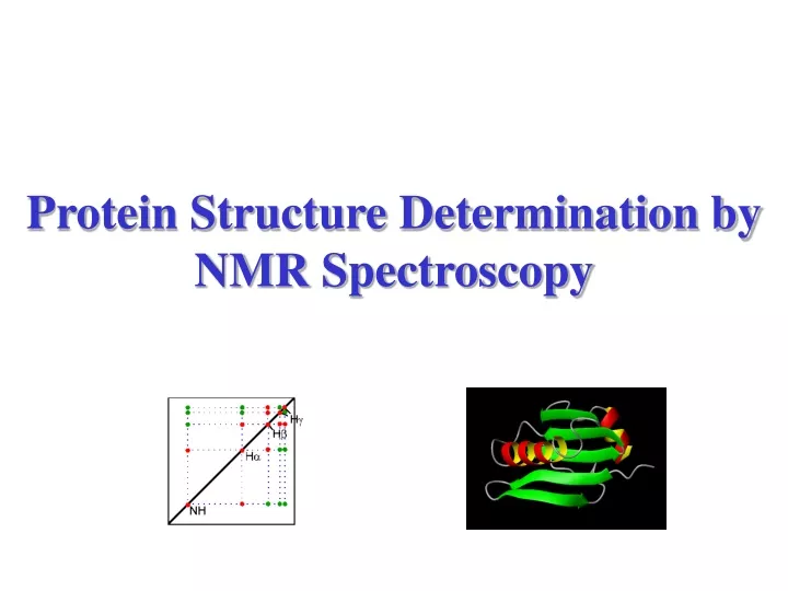
Protein Structure Determination by NMR Spectroscopy. 1D Spectrum. amide/ aromatic/ region. alpha region. methyl region. 0.015 M glucagon 360 MHz. 0.01 M Inhibitor K , 360 MHz. 2D J-resolved. COSY 45. COSY 90. COSY DQF. TOCSY. COSY L.R. TOCSY.

E N D
1D Spectrum amide/ aromatic/ region alpha region methyl region
2D J-resolved COSY 45.COSY 90.COSY DQF.TOCSY. COSY L.R.TOCSY Relayed COSY. (with one homonuclear relayed). NOESY.ROESY.
Protein Structure Determined by NMR 1D NMR NOESY TOCSY DQF-COSY Peak Assignment Distance Constraint Dihedral angle Constraint Structural Simulation
Peak Assignment Strategies • Stage 1 Spin System Identification • Stage 2 Sequence-Specific Assignment
In the COSY experiment, magnetization is transferred by scalar coupling. Protons that are more than three chemical bonds apart give no cross signal because the 4J coupling constants are close to 0. Only signals of protons which are two or three bonds apart are visible in a COSY spectrum (red signals). The cross signals between HN and Halpha protons are of special importance because the phi torsion angle of the protein backbone can be derived from the 3J coupling constant between them The TOCSY experiment correlates all protons of a spin system. Therefore, not only the red signals are visible (which also appear in a COSY spectrum) but also additional signals (green) which originate from the interaction of all protons of a spin system that are not directly connected via three chemical bonds. Thus a characteristic pattern of signals results for each amino acid from which the amino acid can be identified
approximate chemical shifts for various groups in a protein • “random coil” means “not having any specific structure” e.g. helix, sheet • measured in unstructured tetrapeptides • notice that none of these values is above 8.8 (except Trp sidechain NH) or below 0.9 • all the amides come between 8 and 8.75 • alphas between 4 and 4.8 • most methyls between 0.9 and 1.4 • some amino acids have identical spin systems and therefore identical signal patterns. They are: cysteine, aspartic acid, phenylalanine, histidine, asparagine, tryptophane and tyrosine ('AMX systems') on the one hand and glutamic acid, glutamine and methionine ('AM(PT)X systems') on the other hand.
Hd Ha He Hb Hg Hd Hg Hb He Ha
the technique of making the spin-system assignments, followed by sequence-specific assignment using unique fragments of sequence, is known as sequential assignment (Wuthrich)
main-chain directed assignment(Englander). This technique does not focus on assigning all the spin systems first. Rather, it focuses on the backbone and links sizable stretches of backbone residues via sequential (i,i+1) nOe’s and other nOe’s that are characteristic of secondary structures. This technique is particularly useful when there is some knowledge of secondary structure beforehand.
Chemical Shift & Protein Secondary Structure For helical conformation NH & a-H move upfield from the r.c. value For b-strand conformation NH & a-H move downfield from the r.c. value Wishart, Sykes & Richards, J. Mol. Biol. 1991, 222,311-333
化學位移差值(Chemical Shift Index;CSI) • 用來判斷蛋白質及胜二級結構之構形傾向,可以顯現與二級結構的關係,計算方法為: C.S. difference = d測量-d無序纏捲
Temperature Coefficient • 溫度每上升1K,backbone NH 質子所感受到周圍環境的不同,所產生的化學位移變化,溫度係數的單位為ppb/K。 • 溫度係數必須與氫氘交換實驗的結果做比較,才可以判斷背骨胺基質子是否有氫鍵存在。 Baxter & Williamson, J. Biomolecular NMR, 1997, 9 ,359-369
Backbone H-D Exchange 2 hour 30 min gp-41 fusion peptide in SDS micelle, 298 K
Backbone H-D Exchange Amide proton resonance region of the 23-mer fusion peptide of HIV-gp41 in SDS micelle pH 5 pH 7
Nuclear Overhauser Effect Distance & NOE Strength Strong 1.8 ~ 2.7 Å; Medium 1.8 ~ 3.5 Å; Weak 1.8 ~ 5.0 Å