Download
1 / 37
370 likes | 379 Views
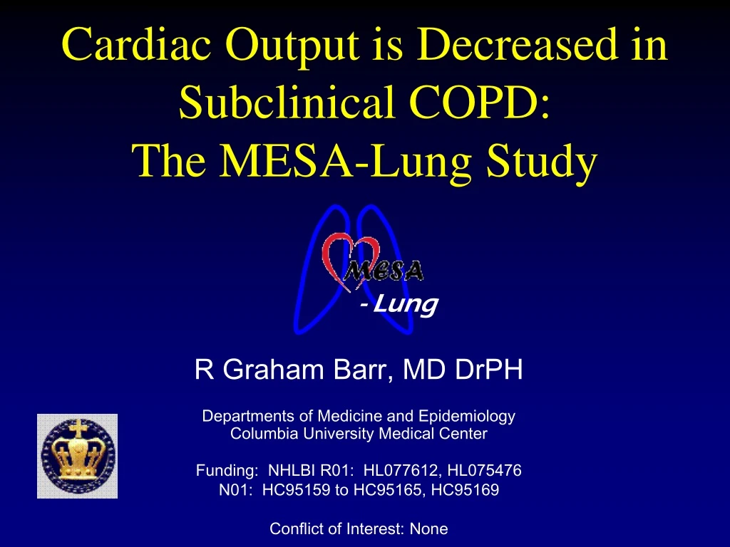
- Lung. Cardiac Output is Decreased in Subclinical COPD: The MESA-Lung Study. R Graham Barr, MD DrPH Departments of Medicine and Epidemiology Columbia University Medical Center Funding: NHLBI R01: HL077612, HL075476 N01: HC95159 to HC95165, HC95169 Conflict of Interest: None. - Lung.

E N D
- Lung Cardiac Output is Decreased in Subclinical COPD: The MESA-Lung Study R Graham Barr, MD DrPH Departments of Medicine and Epidemiology Columbia University Medical Center Funding: NHLBI R01: HL077612, HL075476 N01: HC95159 to HC95165, HC95169 Conflict of Interest: None
- Lung Obstructive Airways Disease CHRONIC BRONCHITIS EMPHYSEMA COPD CHRONIC AIRFLOW OBSTRUCTION ASTHMA
- Lung Death Rate from COPD Doubled1970 - 2002 Jemal 2005, JAMA
- Lung Unexplained Variance in COPD 25 20 15 10 5 0 Non-smoker 1–20 pack-years 21–40 pack-years 41–60 pack-years 61+ pack-years Percentage with abnormal FEV1 Mild Moderate Severe Category of COPD
Early Vascular Hypothesis of Emphysema • Changes in the local vascular milieu modulate alveolar destruction and inflammation that result in emphysema and COPD Leibow, Am Rev Respir Dis, 1959
- Lung COPD “the Blue Bloater” Obese, phlegmy Obstructive spirometry Hypoxemia Pulmonary Hypertension Right Heart Failure Rennard, NEJM, 2004
- Lung Vascular Hypothesis of COPD • During early fetal development, vasculature develops around airways • In late gestation, the capillaries cause epithelial differentiation into type I and II pneumocytes and alveolar formation Control Anti-VEGF van Tuyl, Am J Physiol Lung Cell Mol Physiol, 2005
Endothelial Cell Damage in COPD ControlCOPD (n = 6) (n = 9) Age, yr 64 ± 6 67 ± 9 FEV1, % pred 101 ± 11 63 ± 12* FVC, % pred 96 ± 9 77 ± 14* FEV1/FVC, % 79 ± 7 62 ± 10 RV, % pred 108 ± 10 144 ± 56 DLCO, % pred 80 ± 7 69 ± 16 PaO2, mm Hg 85 ± 6 73 ± 11* PaCO2, mm Hg 36 ± 2 36 ± 2 Peinado, AJRCCM, 2006
Endothelial Hypothesis of COPD Smoking Endothelial dysfunction Endothelial apoptosis Pulmonary Hypertension Alveolar Destruction (Emphysema ↓LV preload and COPD) (↓Cardiac Output) FMD, VEGF, Ceramide, S1P
- Lung Hypothesis • Cardiac output is reduced with airway obstruction (low FEV1/FVC ratio) • The reduction is linear across the distribution of FEV1/FVC ratio • The reduction is greater in smokers than never smokers
- Lung Cardiac Output SV = LVEDV – LVESV CO = HR * SV Estimate of left and right CO Exam 1 cardiac MRI Courtesy: D Bluemke, MD PhD
- Lung Methods – MESA-Lung • MESA-Lung recruited 3,808 participants • Random sample of Exam 3-4 with flow-mediated dilation measures + genetics consent • Oversampled minority race/ethnicity • Spirometry (prebronchodilator) following 1987 American Thoracic Society (ATS) recommendations • Lung density measures on cardiac scans
- Lung Methods – Current Analysis • Current analyses preliminary • Analysis dataset of 2,833, excluding: • Spirometry Quality ‘F’ = 10 • Restrictive Ventilatory Defect = 100
- Lung Methods – Statistics • Pearson correlation coefficients • Generalised additive models with loess smoothers • (S) for CO and main confounders • FEV1/FVC = β0 + S1(CO) + β1(age) + S2(packyr) + … + ε • Nonlinearity of relationships assessed with –2 log likelihood test of nested models • Multivariate interaction with smoking status
- Lung Results – Demographics
- Lung Lung Function and CT Density *n=2024
- Lung Cardiac Output and FEV1/FVC Correlations – overall (n=2,593) r = 0.11, p<0.001 • Current smokers (n=297) r = 0.21, p<0.001 • Former smokers (n=878) r = 0.11, p=0.002 • Never smokers (n=1,436) r = 0.10, p<0.001
- Lung CO and FEV1/FVC Age-adjusted ΔFEV1/FVC ratio Plinear = 0.003 Pnon-linearity = 0.22 Pcurve = 0.22 Cardiac Output N=2,593
- Lung CO and FEV1/FVC Multivariate Interaction CO*smoking P=0.001 ΔFEV1/FVC ratio Plinear = 0.0004 Pnon-linearity = 0.28 Cardiac Output Adjusted for age, sex, race/ethnicity, education, smoking status, packyears, cigars, asthma, hayfever family hx emphysema, dust exposure, BMI, height, DM, FPG, HTN, SBP, DBP, CRP, BSA, LVEF
- Lung Multivariate – Current Smokers Multivariate ΔFEV1/FVC ratio Plinear = 0.0003 Pnon-linearity = 0.53 Cardiac Output Adjusted for age, sex, race/ethnicity, education, smoking status, packyears, cigars, asthma, hayfever family hx emphysema, dust exposure, BMI, height, DM, FPG, HTN, SBP, DBP, CRP, BSA, LVEF
- Lung Multivariate – Past Smokers Multivariate ΔFEV1/FVC ratio Plinear = 0.008 Pnon-linearity = 0.20 Cardiac Output Adjusted for age, sex, race/ethnicity, education, smoking status, packyears, cigars, asthma, hayfever family hx emphysema, dust exposure, BMI, height, DM, FPG, HTN, SBP, DBP, CRP, BSA, LVEF
- Lung Multivariate – Never Smokers Multivariate ΔFEV1/FVC ratio Plinear =0.64 Pnon-linearity = 0.51 Cardiac Output Adjusted for age, sex, race/ethnicity, education, smoking status, packyears, cigars, asthma, hayfever family hx emphysema, dust exposure, BMI, height, DM, FPG, HTN, SBP, DBP, CRP, BSA, LVEF
- Lung Limitations • Preliminary analysis of partial dataset • No post-bronchodilator measures • Lacking RV measures • MESA-RV (R01 HL086719, Kawut) • Possible incomplete adjustment for other confounders
- Lung Conclusions • Cardiac output is reduced early in COPD • Suggests subclinical pulmonary hypertension early in COPD • May be related to early endothelial dysfunction in COPD
- Lung Acknowledgements MESA-Lung Investigators Jeff Carr, MD MS Wake Forest Robert Detrano, MD PhD UCLA-Harbor Paul Enright, MD University of Arizona John Hankinson, PhD Karen Hinckley, MS University of Washington Eric Hoffman, PhD University of Iowa Rui Jiang, MD DrPH Columbia Steve Kawut, MD MPH Columbia Richard Kronmal, PhD University of Washington Kiang Liu, PhD Northwestern Naresh Punjabi Johns Hopkins University Dan Rabinowitz, PhD Columbia Eyal Shahar, MD University of Arizona Lewis Smith, MD Northwestern Russell Tracy, PhD University of Vermont Karol Watson, MD UCLA
- Lung Right Ventricular Ejection Fraction Age-adjusted Multivariate * RVEF (%) P trend = 0.75 P trend = 0.68 None Mild Moderate Severe+ COPD None Mild Moderate Severe+ COPD n = 210 * Adjusted for age, gender, race/ethnicity, LVEF
- Lung SV and FEV1/FVC Multivariate ΔFEV1/FVC ratio Plinear < 0.0001 Pnon-linearity = 0.20 Stroke Volume Adjusted for age, gender, race/ethnicity, education, smoking status, packyears, BMI, diabetes, FPG, hypertension, systolic & diastolic blood pressure, LDL, HDL, CRP, pulse rate, body surface area, LVEF
- Lung Multivariate – Current Smokers Multivariate * ΔStroke volume (mL) Plinear < 0.0001 Pnon-linearity = 0.27 FEV1/FVC ratio * Adjusted for age, gender, race/ethnicity, education, smoking status, packyears, BMI, diabetes, FPG, hypertension, systolic & diastolic blood pressure, LDL, HDL, CRP, pulse rate, BSA, LVEF
- Lung Multivariate – Past Smokers Multivariate * ΔStroke volume (mL) Plinear = 0.0003 Pnon-linearity = 0.30 FEV1/FVC ratio * Adjusted for age, gender, race/ethnicity, education, smoking status, packyears, BMI, diabetes, FPG, hypertension, systolic & diastolic blood pressure, LDL, HDL, CRP, pulse rate, BSA, LVEF
- Lung Multivariate – Never Smokers Multivariate * ΔStroke volume (mL) Plinear = 0.74 Pnon-linearity = 0.66 FEV1/FVC ratio * Adjusted for age, gender, race/ethnicity, education, smoking status, packyears, BMI, diabetes, FPG, hypertension, systolic & diastolic blood pressure, LDL, HDL, CRP, pulse rate, BSA, LVEF
- Lung Cardiovascular Risk Profile
- Lung Left Ventricular Ejection Fraction Age-adjusted Multivariate * LVEF (%) P trend = 0.002 P trend = 0.40 None Mild Moderate Severe+ COPD None Mild Moderate Severe+ COPD * Adjusted for age, gender, race/ethnicity, education, smoking status, packyears, BMI, diabetes, FPG, hypertension, systolic & diastolic blood pressure, LDL, HDL, CRP n = 1,982
- Lung Stroke Volume Age-adjusted Multivariate * Stroke volume (mL) P trend = 0.27 P trend = 0.0004 None Mild Moderate Severe+ COPD None Mild Moderate Severe+ COPD * Adjusted for age, gender, race/ethnicity, education, smoking status, packyears, BMI, diabetes, FPG, hypertension, systolic & diastolic blood pressure, LDL, HDL, CRP, pulse rate, body surface area n = 1,982
- Lung Cardiac Output Age-adjusted Multivariate * Cardiac output (L/min) P trend = 0.09 P trend = 0.0005 None Mild Moderate Severe+ COPD None Mild Moderate Severe+ COPD * Adjusted for age, gender, race/ethnicity, education, smoking status, packyears, BMI, diabetes, FPG, hypertension, systolic & diastolic blood pressure, LDL, HDL, CRP, pulse rate, body surface area n = 1,974
- Lung Spirometry – Quality Factors FVC FEV1 Women Men
- Lung Methods – Spirometry QF • Acceptable Curve1 • Satisfactory start of test • Satisfactory end of test (EOT) – (ET > 6s and 1-s plateau) • No cough during first second • No cough, glottis closure, leak, obstruction, extra breath • Quality Factors • A = 3 acceptable curves, 2 largest within 100 ml (+good EOT) • B = 2 acceptable curves, 2 largest within 150 ml (+good EOT) • C = 2 acceptable curves, 2 largest within 250 ml (+ET > 6s) • D = 1 acceptable curve • F = no acceptable curves 1Miller, MR et al. Standardization of Spirometry, Eur Respir J 2005;26:319-338