Download
1 / 66
800 likes | 1.94k Views
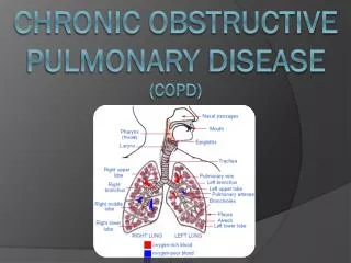
Chronic Obstructive Lung Disease (COPD ) Chronic Bronchitis & Emphysema. LUNG STRUCTURE. CHRONIC OBSTRUCTIVE PULMONARY DISEASE (COPD) . Definition COPD is a chronic , slowly progressive disorder characterised by airflow obstruction.

E N D
Chronic Obstructive Lung Disease (COPD ) Chronic Bronchitis & Emphysema
CHRONIC OBSTRUCTIVE PULMONARY DISEASE (COPD) Definition COPD is a chronic , slowly progressive disorder characterised by airflow obstruction. (FEV1 is less than 80% of the predicted value and FEV1/FVC < 70%. ) which does not change markedly over several months. The impairement of lung function is largely fixed (irreversible) but may be partially reversible by bronchodilator therapy. COPD is unlike asthma, is not fully reversible
Chronic bronchitis Is defined when a cough and sputum occur on most days for at least 3 consecutive months for at least 2 successive years ( provided other causes of cough had been excluded). • The 'blue bloaters( Chronic bronchitis ) is characterized by chronic productive cough, likely to be heavy ( obese) and cyanotic & develop hypercapnia earlier and may develop oedema and secondary polycythaemia.
Emphysema Referrred to the pathological process of a permanent destructive enlargement of the airspaces distal to the terminal bronchioles. . • The 'pink puffers ( Emphysema) is characterized by chronic cough , are typically thin and breathless, and maintain a normal PaCO2 ( noncyanotic) at rest until the late stage of disease. have prominent use of accessory muscles .
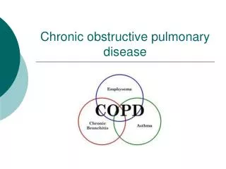
Although pure form of Chronic bronchitis & Emphysema do exist, there is cosiderable overlap in the vast majority of patients. ( COPD predominantely Chronic bronchitis or • COPD predominantely Emphysema ). • In practice, these phenotypes often overlap.
Aetiology of COPD • RISK FACTORS FOR DEVELOPMENT OF COPD • Exposures • Tobacco smoking. • Occupation-coal miners . • Outdoor and indoor air pollution • Low birth weight-may reduce maximally attained lung function in young adult life • Lung growth-insults including childhood infections or maternal smoking may affect growth of lung during childhood, resulting in a lower maximally attained lung function in adult life • Infections-recurrent infection may accelerate decline in FEV1. Persistence of adenovirus in lung tissue may alter local inflammatory response predisposing to lung damage. HIV infection associated with emphysema Host factors • Genetic factors-α1-antiproteinasedeficiency • Airway hyper-reactivity
Aetiology of COPD • A variety of factors appear to increase the risk of developing COPD,but the single most important cause is cigarette smoking. • Smoking cause its effect by inducing persistent airway inflammation & causing a direct imbalance in oxidant\ antioxidant capacity & proteinase/antiproteinase load in the lungs • Only 15% of smokers likely to develop clinically significant COPD & there is a familial risks associated with the development of COPD. • Stopping smoking slows the average rate of the decline in FEV1 from 50 – 70 ml/ year to 30 ml/year (i.e. equal to non-smokers ). • Susceptibility to cigarette smoke varies but both the dose and duration of smoking appear to be important, and it is unusual to develop COPD with less than 10 pack years (1 pack year = 20 cigarettes/day/year). • Alpha 1-antitrypsin deficiency can cause emphysema in non-smokers but this risk is increased dramatically in enzyme-deficient patient who smoke.1–2% of COPD patients are found to have severe 1AT deficiency as a contributing cause of COPD,
If people stop smoking, receive early diagnosis and the right care, COPD’s progression can be slowed down, enabling people to live healthy and active lives for longer
COPD is largely a preventable disease, approximately 80% of cases are attributable to smoking. • Occupational and environmental factors account for approximately 15%, and there is a genetic element in a small number of cases.
COPD will rise from the sixth to the third most common cause of death worldwide by 2020. • COPD is the fourth leading cause of death • Leading causes of death: • Heartdisease • Cancer • Cerebrovasculardisease (stroke) • Respiratorydiseases (COPD) • Accidents • Pneumonia and influenza • Diabetes • Suicide • Nephritis • Chronicliverdisease • All other causes of death
Pathogenesis • Tobacco smoking is the main risk factor for COPD, although other inhaled noxious particles and gases may contribute. • In addition to inflammation, an imbalance of proteinases and antiproteinases in the lungs, and oxidative stress are also important in the pathogenesis of COPD. • Pathophysiology • The different pathogenic mechanisms produce the pathological changes which, in turn, give rise to the physiological abnormalities in COPD: • mucous hypersecretion and ciliary dysfunction, • airflow limitation and hyperinflation, • gas exchange abnormalities, • pulmonary hypertension, • systemic effects.
Pathophysiology • COPD has both pulmonary and systemic components . • An enlargement of mucus-secreting glands and an increased number of goblet cells in the larger airways contribute to enhanced secretion of airway mucus that manifests as chronic bronchitis. • Loss of elastic tissue surrounding the smaller airways, accompanied by inflammation and fibrosis in the airway wall and mucus accumulation within the airway lumen, results in airflow limitation, further increased by enhanced cholinergic tone. • Premature airway closure leads to gas trapping and hyperinflation, which in turn decrease pulmonary and chest wall compliance. • During exercise, the time available for expiration shortens, resulting in progressive hyperinflation. • The work of breathing is therefore markedly increased, first on exercise but then, as the disease advances, at rest.
In the alveolar capillary units the unopposed action of proteases and oxidants results in destruction of the alveoli and the appearance of emphysema . Emphysema may be classified by the pattern of the enlarged airspaces: centriacinar, panacinar and periacinar. Bullaeform in some individuals. In COPD there is often "air trapping" (increased residual volume and increased ratio of residual volume to total lung capacity) and progressive hyperinflation (increased total lung capacity) late in the disease. COPD may results in impaired gas exchange and respiratory failure.
Inflammatory cells produce elastase • Destroys connective tissue of alveolar walls • Alpha-1 anti-trypsin (or alpha-1 protease inhibitor) is a protein produced by the liver that circulates in the blood and limits the action of elastase
Systemic effects of COPD • Muscular weakness (cellular changes in skeletal muscles ). • Impaired salt & water excretion leading to peripheral oedema. • Altered fat metabolism contributing to weight loss • Increased prevalence of osteoporosis. • Increased circulating inflammatory markers.
Pathogenesis of COPD NOXIOUS AGENT(tobacco smoke, pollutants, occupational agent) COPD Genetic factors Respiratory infection Other
Clinical features of COPD • Clinical features COPD should be suspected in any patient over the age of 40 years who presents with symptoms of persistent cough sputumproduction breathlessness. Many patients have such symptoms for months or years before seeking medical attention • Depending on the presentation important differential diagnoses include asthma, tuberculosis, bronchiectasis and congestive cardiac failure. • Chronic severe asthma may be difficult to distinguish from COPD.
Clinical features of COPD • Cough is usually the first symptom but seldom prompts the patient to consult a doctor. • It is characteristically accompanied by small amounts of mucoid sputum. • Chronic bronchitis is formally defined when a cough and sputum occur on most days for at least 3 consecutive months for at least 2 successive years. • Haemoptysismay complicate exacerbations of COPD but should not be attributed to COPD without thorough investigation. • Breathlessnessusually heralds the first presentation to the health professional. • In advanced disease, enquiry should be made as to the presence of oedema(which may be seen for the first time during an exacerbation) and • morning headachesindicative of hypercapnia.
Physical signs • The presence of pitting oedema should be documented and the body mass index (BMI) recorded. • Crackles may accompany infection but if persistent raise the possibility of bronchiectasis. • Finger clubbing is not consistent with COPD and should alert the physician to potentially more serious pathology. • Two classical phenotypes have been described: 'pink puffers' and 'blue bloaters'. • The 'pink puffers ( Emphysema) are typically thin and breathless, and maintain a normal PaCO2 ( noncyanotic) at rest until the late stage of disease. have prominent use of accessory muscles, • The 'blue bloaters( Chronic bronchitis ) likely to be heavy and cyanotic & develop hypercapnia earlier and may develop oedema and secondary polycythaemia. • In practice, these phenotypes often overlap.
Clinical Abnormalities in patients with advanced Airway obstruction ( COPD ) • A reduction in the length of the trachea palpable above the sternal notch. • Tracheal descent during inspiration(tracheal tug) • Contraction of the sternomastoid and scalene muscles on inspiration • Excavation of the suprasternal and supraclavicularfossaeduring inspiration,together with indrawing of the costal margins and intercostal spaces. • Loss of weight (often stimulates unnecessary investigation) • Pursed lip breathing– physiological response to decrease air trapping . • Central cyanosis • Flapping tremor and bounding pulse(due to hypercapnia)
Increased antero-posterior diameter of the chest relative to the lateral diameter (signs of hyperinflation include a barrel chest ). • decreased tactile vocal fremitus. • hyperresonant percussion note • loss of cardiac & hepatic dullness • decreased breath sounds; prolonged expiratory phase and expiratory wheezing. (Rhonchi,especially on forced expiration ). • Peripheral oedemawhich may indicate corpulmonale • Raised JVP, right ventricular heave, loud pulmonary second sound, tricuspid regurgitation.
Advanced disease may be accompanied by systemic wasting, with significant weight loss, bitemporal wasting, and diffuse loss of subcutaneous adipose tissue. This syndrome has been associated with both inadequate oral intake and elevated levels of inflammatory cytokines (TNF-). Such wasting is an independent poor prognostic factor in COPD. • Clubbingof the digits is not a sign of COPD, and its presence should alert the clinician to initiate an investigation for causes of clubbing. In this population, the development of lung cancer is the most likely explanation for newly developed clubbing
ASSESSMENT OF SEVERITY OF AIRFLOW OBSTRUCTION ACCORDING TO FEV1 ( classification of COPD according to the severity ) • SeverityFEV1 • Mild 50-80% predicted • Moderate 30-49% predicted • Severe < 30% predicted
Complications of COPD • Pulmonary bullae: Are thin-walled airspaces created by rupture of alveolar walls. They may be single or multiple , large or small & tend to be situated subpleurally , Rupture of subpleuralbullae may cause pneumothorax,& occationallybullae increase in size , compress functioning lung tissue & further embarrass pulmonary ventilation. Respiratory failure & corpulmonaleare generally late complications in COPD patients.
COPD-Investigations Pulmonary function tests The diagnosis of COPD requires objective demonstration of airflow obstruction by spirometry and is established when FEV1 is less than 80% of the predicted value and accompanied by FEV1/FVC < 70% normal FEV1 exclude the diagnosis of COPD Reversability test is necessory to detect asthmatic cases. Lung volumes show an increase in TLC & RV due to gas trapping The carbone monoxide transfer factor & coefficient are markedly reduced in patients with sever emphysema component
Diagnosis of COPD EXPOSURE TO RISK FACTORS SYMPTOMS cough tobacco sputum occupation dyspnea indoor/outdoor pollution è SPIROMETRY
Measurement of arterial blood gases • should be performed in all patients with sever COPD ( FEV1 less than 40% ) • Alveolar underventillation causes a fall in paO2 & often a perminant increase inpaCO2. • Pulse oximetry may prompt referral for a domiciliary oxygen assessment if less than 93%.
Imaging 1- chest X-ray • In moderate to severe COPD the chest X-ray typically shows : • hypertranslucent lung fields . • with disorganisation of vasculature . • low flat diaphragm . • prominent pulmonary artery shadows at both hila. • Bullae may also be observed. chest X-ray is essential to identify alternative diagnoses such as cardiac failure, other complications of smoking such as lung cancer.
2- scan of chest • can be used to quantify the extent & distribution of emphysema & for the assessment of bullous emphysema & the potential for lung volume reduction surgery or lung transplantation. • CT is likely to play an increasing role in the assessment of COPD as it allows the detection, characterisation and quantification of emphysema and is more sensitive than the chest X-ray at detecting bullae • Patients with with α1-antitrypsin deficiency typically display basal disease , compared with the predominantly apical disease seen in smokers with normal α1-antitrypsin level
Haematology • Polycythemia (secondary Polycythemia)may develop, but should not be assumed to be secondary without measurement of paO2 • Venesection may be considered if the haematocrit is above 0.55 • In younger patients with predominantly basal emphysema, α1-antitrypsin level should be assayed.
Management of COPD Reduction of bronchial irritation: Smoking cessation advise and assist the patient toward smoking cessation.. Cessation is difficult but highly rewarding and remains the only intervention proven to decelerate the decline in FEV1. Dusty & smoke laden atmospheres should be avoided reduction of occupational exposure( this may involve a change of occupation).
Reduction of total personal exposure to tobacco smoke, occupational dusts and chemicals, and indoor and outdoor air pollutants are important goals to prevent the onset and progression of COPD. • Smoking cessation is the single most effective - and cost effective - intervention to reduce the risk of developing COPD and stop its progression.
Bronchodilators Bronchodilator therapy is central to the management of breathlessness in patients with COPD. The principal bronchodilator treatments are beta2-agonists,anticholinergics, theophylline, and a combination of these drugs The inhaled route is preferred. Short-acting bronchodilators may be used for patients with mild disease but longer-acting bronchodilators are more appropriate for patients with moderate to severe disease. Oral bronchodilator therapy may be used in patients who cannot use inhaled devices efficiently. .
Bronchodilator therapy with regular inhaled anticholinergic agents & short acting B2 agonists taken as required provides useful symptomatic relief in the majority of patients. The choice between beta2-agonist, anticholinergic, theophylline, or combination therapy depends on availability and individual response in terms of symptom relief and side effects. Combining bronchodilators may improve efficacy and decrease the risk of side effects compared to increasing the dose of a single bronchodilator
Corticosteroids Inhaled corticosteroids (ICS) reduce the frequency and severity of exacerbations. They are currently recommended in patients with severe disease (FEV1 < 50 % ) who report two or more exacerbations requiring antibiotics or oral steroids per year. The combination of ICS with long-acting β 2-agonists produces further improvement in breathlessness and reduces the frequency and severity of exacerbations. Oral corticosteroids are useful during exacerbations but maintenance therapy contributes to osteoporosis and impaired skeletal muscle function and should be avoided. .
Other measures Exercise should be encouraged & outpatient based pulmonary rehabilitation programmes, while not affecting the FEV1, can improve exercise performance & reduce breathlessness. Obesity , poor nutrition , depression & social isolation should be managed. Sedatives & opiate-based analgesic preparations are contraindicated.
LONG-TERM DOMICILIARY OXYGEN THERAPY • In patients with COPD Long term low – concentration oxygen therapy ( 2 litres/min by nasal canulae for ≥ 15 hrs/day intermittently) . • It reduces secondary polycythaemia ,decrease pulmonary hypertension & improves neuropsychological health & most importantly prolong life in hypoxaemic COPD patients. • The aim of therapy is to increase the PaO2 to at least 8 kPa (60 mmHg) or SaO2 at least 90% . • High concentrations of oxygen may cause respiratory depression and worsening acidosis .
PRESCRIPTION OF LONG-TERM OXYGEN THERAPY (LTOT) IN COPD Arterial blood gases measured in clinically stable patients on optimal medical therapy on at least two occasions 3 weeks apart. PaO2≤ 7.3 kPa (55 mmHg) irrespective of PaCO2 and FEV1 ≤ 1.5 litres. PaO27.3 - 8 k Pa (55-60 mmHg) plus pulmonary hypertension, peripheral oedema or nocturnal hypoxaemia Patient stopped smoking . Use at least 15 hours/day at 2-4 litres/min to achieve a PaO2 > 8 kPa (60 mmHg) without unacceptable rise in PaCO2