Download
1 / 7
80 likes | 345 Views
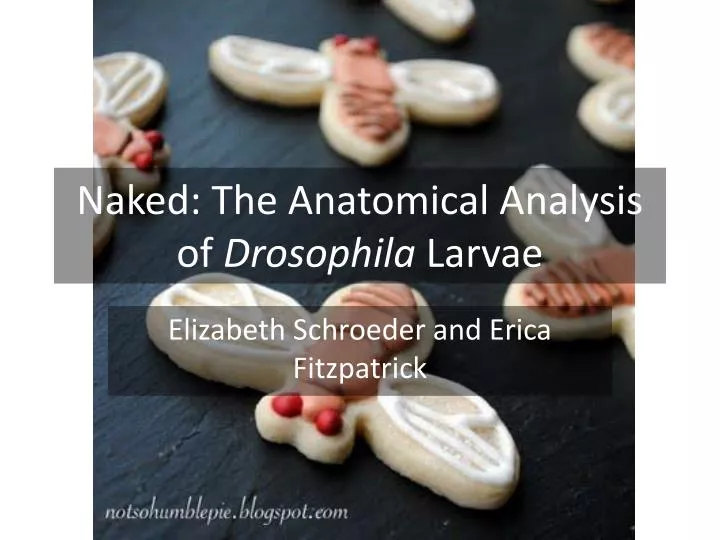
Naked: The Anatomical Analysis of Drosophila Larvae. Elizabeth Schroeder and Erica Fitzpatrick. The entire anatomy of a drosophila larva. http://www.morgellonsuk.org.uk/images/fly_larvae/flylarvae_anatomy.gif.

E N D
Naked: The Anatomical Analysis of Drosophila Larvae Elizabeth Schroeder and Erica Fitzpatrick
The entire anatomy of a drosophila larva http://www.morgellonsuk.org.uk/images/fly_larvae/flylarvae_anatomy.gif Our image of a drosophila larva shows the muscles near the mouth hooks and the gut stained blue
Spiracles http://ny-image2.etsy.com/il_fullxfull.82528390.jpg This is the slightly disfigured head of a larva. The spiracles are stained blue on either side of the head.
Spiracle Trachea http://www.morgellonsuk.org.uk/images/fly_larvae/Drosophila_larvae_spiracle_mouth.jpg This is another image of the head of a larva. The stained blue parts belong to the spiracles.
Salivary Gland Another image of a disfigured head. The blue triangular structure on the bottom left is the salivary gland http://www.uncg.edu/bio/facilities/confocal/Images/Drosophila%20melanogaster%20salivary%20gland.jpg
Midgut http://lemaitrelab.epfl.ch/files/content/sites/lemaitrelab/files/users/182725/public/Fig4.JPG This image shows section of the larva’s gut. The blue section is the midgut. (Don’t mind the bubble)
MidgutImaginal Islands A zoomed-in section of the gut. The little blue dots are called the midgutimaginal islands, premature cells located in the midgut. http://www.uncg.edu/bio/facilities/confocal/Images/DrosophilaMidGut.jpg