Download
1 / 18
190 likes | 294 Views
Gel Electrophoresis of DNA. Molecular Genetics Presentation by: Nana Sugma Mulyana Febrina Anggraini Ginting Faizal Dony Rifai Nor Aviva. Acknowledgements. Thanks go to Craig Millar, School of Biological Science, University of Auckland Compiled by Linda Macdonald

E N D
Gel Electrophoresis of DNA • Molecular Genetics Presentation by: • Nana Sugma Mulyana • Febrina Anggraini Ginting • Faizal Dony Rifai • Nor Aviva
Acknowledgements Thanks go to Craig Millar, School of Biological Science, University of Auckland Compiled by Linda Macdonald For NCEA Biology A.S. 3.6 With support from the Royal Society Science, Mathematics &Technology Teacher Fellowship Scheme
What is Gel Electrophoresis? • Electro = flow of electricity, phoresis, from the Greek = to carry across • A gel is a colloid, a suspension of tiny particles in a medium, occurring in a solid form, like gelatin • Gel electrophoresis refers to the separation of charged particles located in a gel when an electric current is applied • Charged particles can include DNA, amino acids, peptides, etc
Why do gel electrophoresis? • When DNA is cut by restriction enzymes, the result is a mix of pieces of DNA of different lengths • It is useful to be able to separate the pieces - I.e. for recovering particular pieces of DNA, for forensic work or for sequencing
What is needed? • Agarose - a polysaccharide made from seaweed. Agarose is dissolved in buffer and heated, then cools to a gelatinous solid with a network of crosslinked molecules • Some gels are made with acrylamide if sharper bands are required
Buffer - in this case TBE • The buffer provides ions in solution to ensure electrical conductivity. • Not only is the agarose dissolved in buffer, but the gel slab is submerged (submarine gel) in buffer after hardening
Also needed are a power supply and a gel chamber • Gel chambers come in a variety of models, from commercial through home-made, and a variety of sizes
How does it work? • DNA is an organic acid, and is negatively charged (remember, DNA for Negative) • When the DNA is exposed to an electrical field, the particles migrate toward the positive electrode • Smaller pieces of DNA can travel further in a given time than larger pieces
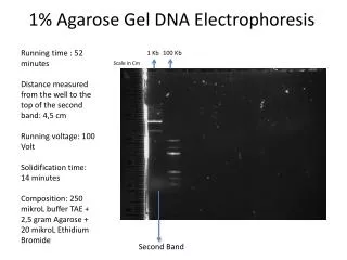
A gel being run Positive electrode Comb Agarose block DNA loaded in wells in the agarose Buffer Black background To make loading wells easier
Steps in running a gel • DNA is prepared by digestion with restriction enzymes • Agarose is made to an appropriate thickness (the higher the % agarose, the slower the big fragments run) and ‘melted’ in the microwave • The gel chamber is set up, the ‘comb’ is inserted • The agarose may have a DNA ‘dye’ added (or it may be stained later). The agarose is poured onto the gel block and cooled
The comb is removed, leaving little ‘wells’ and buffer is poured over the gel to cover it completely • The DNA samples are mixed with a dense loading dye so they sink into their wells and can be seen
The DNA samples are put in the wells with a micropipette. • Micropipettes have disposable tips and can accurately measure 1/1,000,000 of a litre
Next? • The power source is turned on and the gel is run. The time of the run depends upon the amount of current and % gel, and requires experimentation • At the end of the run the gel is removed (it is actually quite stiff) • The gel is then visualized - UV light causes the bands of DNA to fluoresce
Animation 1 Simple 2 More…
A gel as seen under UV light - some samples had 2 fragments of DNA, while others had none or one
More…… • Many samples can be run on one gel- but it is important to keep track • Most gels have one lane as a ‘DNA ladder’ - DNA fragments of known size are used for comparison
Still more…. • The DNA band of interest can be cut out of the gel and the DNA extracted - • Or DNA can be removed from the gel by Southern Blotting
References www.biotech.iastate.edu/publication/ppt-presentations Kreuzer, H., Massey, A., 2001, Recombinant DNA and Biotechnology,2nd ed. ASM Press, Washington Turner, P.C., et al, 1997, Instant Notes in Molecular Biology, Bios, Oxford Photos - L. D. Macdonald, 2003