Download
1 / 24
240 likes | 264 Views
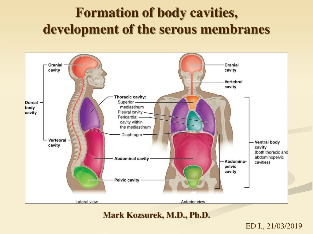
Formation of body cavities , development of the serous membranes. Mark Kozsurek, M.D., Ph.D. ED I., 21/03/2019. Appearance of the common pericardio-pleuro-peritoneal cavity . Isolation of the pleural and peritoneal cavities: the formation of the diaphragm .

E N D
Formation of body cavities, development of theserousmembranes Mark Kozsurek, M.D., Ph.D. ED I., 21/03/2019
Appearance of thecommonpericardio-pleuro-peritonealcavity. • Isolation of the pleural and peritoneal cavities: the formation of the diaphragm. • Isolation of the pericardiac and pleural cavities. • Furtherdevelopment of thepericardium, pleura and peritoneum.
A) Appearance of thecommonpericardio-pleuro-peritonealcavity Back tothe 2nd week: bilaminarembryonicdiskcomposed of epiblasts and hypoblasts During the 3rd week: proliferation of epiblastsresults in gastrulation
Gastrulation:junctionsamongepiblastsbecomelooser and duetothecollisionattheprimitivenode and primitivestreakepiblasts sink belowtheiroriginallayer. Some of themreplacethehypoblasts and differentiateintotheultimateendoderm, whileothers, themesoblasts, form a third, middlelayer, theintraembryonicmesoderm. Epiblastremaining in theiroriginallayerwillformtheectoderm.
Initiallytheintraembryonicmesoderm is just a homogenousmass of tissueonthetwosides of thenotochord. Later a mediolateraldifferentiationgivesarisetothesomites, intermediatemesoderm and thelateralplate. Withinthelateralplatecavitiesform and mergeresulting in theisolation of thesomatic (parietal) and splanchnic (visceral) layer of lateralplatemesoderm.
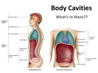
Somatic and splanchniclayers of thelateralplatemesodermenclosethehorse-shoeshaped (orsimply „U”-shaped) intraembryoniccoelom, whichposteriorlyonthelateralsides of theembyocommunicateswiththeextraembryoniccoelom. Withthefurthergrowth of theembryotheintraembryoniccoelomwilldilateanteriorly (thecurve of U - futurepericardium), theproximallimb of U remainsnarrow (pericardioperitonealcanal - thefuturepleuralcavity), whilethevolume of thedistallimb of U alsoincreases (futureperitonealcavity). Oropharyngealmembrane
Isolatedintraembryoniccoelom futurepericardiaccavity Oropharyngealmembrane pericardio-peritoneal canals (future pleural cavities) future peritoneal cavity connectionsbetweentheintra- and extraembryoniccavities
Oropharyngealmembrane A A B B In section B it is wellseenthattheextraembryonicmesoderm is continuouswiththeintraembryonicone, extra- and intraembryoniccoelomscommunicate.
During the 4th week: folding of theembryo has thehighestimpactonthefurterdevelopment of theintraembryoniccavity: heartprimordiummovesontotheventralside of theembryo, septumtransversumturnsintothetransverseplanecaudaltotheheart.
As a result of folding of theembryotheleft and rightlimbs of theintraembryoniccoelomgetclosertothemidline and toeachother and atthesametime, followingthemovements of theheartprimordium, thedilatedcurve of the U-shapedtunnelturnsontotheventralside of theembryo. Butatthisstagethefurtherpericardiac, pleural and peritonealcavitiesarenotisolatedfromoneother (commonpericardiaco-pleuro-peritonealspace).
B) Isolation of thepleural and peritonealcavities: theformation of thediaphragm Notethecentraltendon and thesternal, costal, lumbarparts of thediaphragm. How is itmade?
The narrowpericardiacoperitonealcanalsarefoundbehindtheseptumtransversum. Therearefourstructurescontributingtothediaphragm: septumtransversum pleuroperitonealmembranes– astheygrowthepericardiaco-peritonealcanalnarrows dorsalmesentery of theesophagus – surroundsthe aorta, theesophagus and the IVC muscularingrowthfrom body wall
septumtransversum esophagealmesentery pleuro- peritoneal folds muscularingrowth Myoblastfrom C3-C5 myotomesmigrateintotheseptumtransversum and taketheirnervewiththemselves (phrenicnerve). Butlowerintercostalnervesalsocontributetotheinnervation of theperiphery of thediaphragmformedbythemuscularingrowth of the body wall.
Congenitaldiaphragmatichernia Incompletefusion of thefour parts. Stomach and/orintestineascendintothethoraciccage and compressthelungs.
C) Isolation of thepericardiac and pleuralcavities Formation of thediaphragmhaveisolatedperitonealcavityfromthepericardiacopleuralspace. Diaphragm C3-C5 commoncardinalvein, phrenicnerve Asthehearttubeloopsitscaudal end withthecommoncardinalveinsmovesupward and thephrenicnervesmigratetowardthemidlineformingthepleuropericardial fold.
Duetothechangingposition of thephrenicnerve and thecommoncardinalvein, thepleuro-pericardialmembraneappears, whichlatercompletlyisolatespleural and pericardiaccavities.
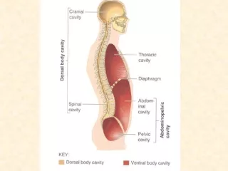
Summary 1.The most anterior part, theloop of theintraembryoniccoelomgivesthepericardialcavityenclosingtheheart. 2. Rostrallimbsof theintraembryoniccoelomdonotunite in themidline and as a result of this, two non-communicating, left and rightpleuralcavitiesdevelop. 3. Caudallimbs of theintraembryoniccoelomremainpairedstructuresanteriorlybuttheymergecaudally. Thisexplainswhytheventralmesentery is onlyobservable in theupperone-third of theabdominalcavity, whilethedorsalmesenteryexistsalongthewholelength of it.
D) Furtherdevelopment of thepericardium Asthehearttubeloops, itsarterious and venousendsapproacheachother. Pericardialreflections (viscerallayer – parietallayertransition) arefoundaroundbothends. Betweenthetworeflectionsthetransverse sinus (T) is found.
E) Furtherdevelopment of thepleura Developinglungsgrowintotheleft and rightpleuralcavities. Transitionbetweenthevisceral and parietallayers (pleuralreflection) surroundstheroot of lung and descentstothediaphragmasthepulmonary ligament. pulmonary ligament
A 3D model of theperitonealcavitydemonstratingthat in theupperone-thirdtheleft and right body cavitiesremainseparated (bothventral and dorsalmesogastrium is seen), butcaudallytheymerge and onlythedorsalmesenterypersists.
right left In theupperone-third of theabdominalcavitytheliver and the spleen developswithintheventral and dorsalmesogastrium, respectively. Astheresult of therotation of theforegut and relatedtransposition of epigastricstructures, thelessersacortheomentalbursaappears. Accordingtothecolourcodethat is derivedfromtheright body cavity (Pleasenote, thatthesidesarenotlabelledcorrectly in Prof. Réthelyi’sFunctionalanatomytextbook).