Download

1 / 59
660 likes | 1.39k Views
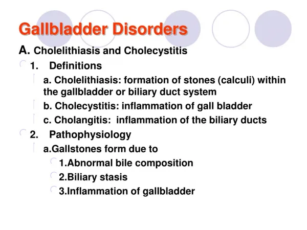
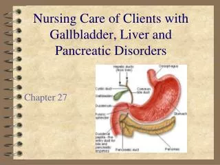
Gallbladder Disorders. A. Cholelithiasis and Cholecystitis 1. Definitions a. Cholelithiasis: formation of stones (calculi) within the gallbladder or biliary duct system b. Cholecystitis: inflammation of gall bladder c. Cholangitis: inflammation of the biliary ducts 2. Pathophysiology

E N D
Gallbladder Disorders A. Cholelithiasis and Cholecystitis • 1. Definitions • a. Cholelithiasis: formation of stones (calculi) within the gallbladder or biliary duct system • b. Cholecystitis: inflammation of gall bladder • c. Cholangitis: inflammation of the biliary ducts • 2. Pathophysiology • a.Gallstones form due to • 1.Abnormal bile composition • 2.Biliary stasis • 3.Inflammation of gallbladder
Gallbladder Disorders • b. Most gallstones are composed primarily of bile (80%); remainder are composed of a mixture of bile components • c. Excess cholesterol in bile is associated with obesity, high-cholesterol diet and drugs that lower cholesterol levels • d. If stones from gallbladder lodge in the cystic duct • 1. There can be reflux of bile into the gallbladder and liver • 2. Gallbladder has increased pressure leading to ischemia and inflammation • 3. Severe ischemia can lead to necrosis of the gall bladder • 4. If the common bile duct is obstructed, pancreatitis can develop
Gallbladder Disorders Risk factors for cholelithiasis • a. Age • b. Family history, also Native Americans and persons of northern European heritage • c. Obesity, hyperlipidemia • d. Females, use of oral contraceptives • e. Conditions which lead to biliary stasis: pregnancy, fasting, prolonged parenteral nutrition • f. Diseases including cirrhosis, ileal disease or resection, sickle-cell anemia, glucose intolerance
Gallbladder Disorders Manifestations of cholelithiasis • a. Many persons are asymptomatic • b. Early symptoms are epigastic fullness after meals or mild distress after eating a fatty meal • c. Biliary colic (if stone is blocking cystic or common bile duct): steady pain in epigastric or RUQ of abdomen lasting up to 5 hours with nausea and vomiting • d. Jaundice may occur if there is obstruction of common bile duct
Gallbladder Disorders Manifestations of acute cholecystitis • a. Episode of biliary colic involving RUQ pain radiating to back, right scapula, or shoulder; the pain may be aggravated by movement, or deep breathing and may last 12 – 18 hours • b. Anorexia, nausea, and vomiting • c. Fever with chills
Gallbladder Disorders Complications of cholecystitis • a. Chronic cholecystitis occurs after repeated attacks of acute cholecystitis; often asymptomatic • b. Empyema: collection of infected fluid within gallbladder • c. Gangrene of gall bladder with perforation leading to peritonitis, abscess formation • d. Pancreatitis, liver damage, intestinal obstruction
Gallbladder Disorders Collaborative Care • a. Treatment depends on the acuity of symptoms and client’s health status • b. Clients experiencing symptoms are usually treated with surgical removal of the stones and gallbladder Diagnostic Tests • a. Serum bilirubin: conjugated bilirubin is elevated with bile duct obstruction • b. CBC reveals elevation in the WBC as with infection and inflammation • c. Serum amylase and lipase are elevated, if obstruction of the common bile duct has caused pancreatitis • d. Ultrasound of gallbladder: identifies presence of gallstones • e. Other tests may include flat plate of the abdomen, oral cholecytogram, gall bladder scan
Gallbladder Disorders Treatment • a. Treatment of choice is laparoscopic cholecystectomy • b. If surgery is inappropriate due to client condition • 1. May attempt to dissolve the gallstones with medications • 2. Medications are costly, long duration • 3. Stones reoccur when treatment is stopped Laparoscopic cholecystectomy • a. Minimally invasive procedure with low risk of complications; required hospital stay< 24 hours. • b. Learning needs of client and family/caregiver include pain control, deep breathing, mobilization, incisional care and nutritional/fluids needs • c. Client is given phone contact for problems
Gallbladder Disorders Some clients require a surgical laparotomy (incision inside the abdomen) to remove gall bladder • a. client will have nasogastric tube in place post-operatively and require several days of hospitalization • b. If exploration of the common bile duct is done with the cholecystectomy, the client may have a T-tube inserted which promotes bile passage to the outside as area heals Clients with cholelithiasis and cholecystitis prior to surgery can avoid future attacks by limiting fat intake Nursing Diagnoses • a. Pain • b. Imbalanced Nutrition: Less than body requirements • c. Risk for Infection
Liver Disorders A. Hepatitis • 1. Definition: inflammation of the liver due to virus, exposure to alcohol, drugs, toxins; may be acute or chronic in nature • 2. Pathophysiology: metabolic functions and bile elimination functions of the liver are disrupted by the inflammation of the liver.
Liver Disorders Viral Hepatitis • 1. Types (causative agents) a. Hepatitis A virus (HAV) Infectious hepatitis • 1. Transmission: fecal-oral route, often contaminated foods, water or direct contact • 2. Contagious through stool up to 2 weeks before symptoms occur; abrupt onset • 3. Benign, self limited; symptoms last up to 2 months
Liver Disorders Hepatitis B virus (HBV) • 1. Transmission: infected blood and body fluids • 2. Liver cells damaged by immune response; increased risk for primary liver cancer; causes acute and chronic hepatitis, fulminant hepatitis and carrier state • 3. High risk to health care workers, injection drug users, men who have sex with men, persons with multiple sexual partners, persons exposed to blood products (hemodialysis clients)
Liver Disorders Hepatitis C virus (HCV) • 1. Transmission: infected blood and body fluids; injection drug use is primary factor • 2. Initial manifestations are mild, nonspecific • 3. Primary worldwide cause of chronic hepatitis, cirrhosis, liver cancer
Liver Disorders Hepatits B-associated delta virus (HDV) • 1. Transmission: infected blood and body fluids; causes infection in people who are also infected with hepatitis B • 2. Causes acute or chronic infection Hepatitis E virus (HEV) • 1. Transmission: fecal-oral route, contaminated water supplies in developing nations; rare in U.S. • 2. Affects young adults; fulminant in pregnant women
Liver Disorders Disease Pattern Associated with hepatitis (all types) • A .Incubation Phase (period after exposure to virus): no symptoms • B Prodromal Phase (preicteric – before jaundice) • 1. “Flu” symptoms: general malaise, anorexia, fatigue, muscle and body aches • 2. Nausea, vomiting, diarrhea, constipation, and mild RUQ abdominal pain • 3. Chills and fever • c.Icteric (jaundiced) Phase • 1 5 – 10 days after prodromal symptoms • 2. Jaundice of the sclera, skin and mucous membranes occurs • 3. Elevation of serum bilirubin • 4. Pruritis • 5. Stool become light brown or clay colored • 6. Urine is brownish colored
Liver Disorders Convalescent Phase • 1. In uncomplicated cases, symptoms improve and spontaneous recovery occurs within 2 weeks of jaundice • 2. Lasts several weeks; continued improvement and liver enzymes improve
Liver Disorders • Chronic Hepatitis • a. Chronic hepatitis: chronic infection from viruses: HBV, HBC, HBD • 1.Few symptoms (fatigue, malaise, hepatomegaly) • 2.Primary cause of cirrhosis, liver, cancer, liver transplants • 3.Liver enzymes are elevated b.Fulminant hepatitis; rapidly progressive disease with liver failure developing within 2 – 3 week of onset of symptoms; rare, but usually due to HBV with HBD infections • c.Toxic hepatitis • 1.Hepatocellular damage results from toxic substances • 2.Includes alcoholic hepatitis, acute toxic reaction or chronic use
Liver Disorders Chronic Hepatitis • a. Chronic hepatitis: chronic infection from viruses: HBV, HBC, HBD • 1. Few symptoms (fatigue, malaise, hepatomegaly) • 2. Primary cause of cirrhosis, liver, cancer, liver transplants • 3. Liver enzymes are elevated • b. Fulminant hepatitis; rapidly progressive disease with liver failure developing within 2 – 3 week of onset of symptoms; rare, but usually due to HBV with HBD infections • c. Toxic hepatitis • 1. Hepatocellular damage results from toxic substances • 2. Includes alcoholic hepatitis, acute toxic reaction or chronic use
Liver Disorders Collaborative Care: Focus is on determination of cause, treatment and support, and prevention future liver damage Diagnostic Tests a. Liver function tests • 1. Alanine aminotransferase (ALT): specific to liver • 2. Aspartate aminotransferase (AST): heart and liver cells • 3. Alkaline phosphatase (ALP): liver and bone cells • 4. Gamma-glutamyltransferase (GGT): present in cell membranes; rises with hepatitis and obstructive biliary disease • 5. Lactic dehydrogenase (LDH): present in many body tissues; isoenzyme, LDH5 is specific to the liver • 6. Serum bilirubin levels: total, conjugated, unconjugated
Liver Disorders b. Lab tests for viral antigens and antibodies associated with types of viral hepatitis c. Liver biopsy: tissue examined to detect changes and make diagnosis • 1. Preparation: signed consent; NPO 4 – 6 hours before • 2. Prothrombin time and platelet count results; may need Vitamin K first to correct • 3. Client voids prior to procedure, supine position • 4. Local anesthetic; client instructed to hold breath during needle insertion • 5. Direct pressure applied to site after sample obtained; client placed on right side to maintain site pressure • 6. Vital signs monitored frequently for 2 hours • 7. No coughing, lifting, straining 1 – 2 weeks afterward
Liver Disorders Medications for prevention of hepatitis • a. Vaccines available for Hepatitis A and B • b. Vaccine for Hepatitis B recommended for high-risk groups • c. Post exposure prophylaxis recommended for household and sexual contacts of persons with HAV or HBV • d. Hepatitis A prophylaxis: single dose of immune globulin within 2 weeks of exposure • e. Hepatitis B prophylaxis: Hepatitis B immune globulin (HBIG) for short-term immunity; HBV vaccine may be given at the same time
Liver Disorders Treatment • a. Medications • 1. Medication for acute hepatitis C: interferon alpha to prevent chronic hepatitis • 2. Chronic Hepatitis B: interferon alpha intramuscular or subcutaneously or lamivudine • 3. Chronic Hepatitis C: interferon alpha with ribavirin (Rebetol) oral antiviral drug
Liver Disorders b. Acute hepatitis treatment • 1. As needed bedrest • 2. Adequate nutrition • 3. Avoid substances toxic to the liver especially alcohol c. Complementary therapies: Milk thistle (silymarin) 8. Nursing Care: Teaching about prevention by stressing • a. Hygiene • b. Handwashing, especially for food handlers • c. Blood and body fluids precautions • d. Vaccines for persons at high risk
Liver Disorders Nursing Diagnoses • a. Risk for Infection • 1.Standard precautions, proper hand washing at all times • 2.Reporting of contagious disease to health department to control spread of disease • b. Fatigue • 1.Scheduling planned rest periods • 2.Gradual increase of activity with improvement • c. Imbalanced Nutrition: Less than body requirements • 1.High caloric diet with adequate carbohydrates • 2.Small frequent meals; nutritional supplements • d. Body Image Disturbance Home care must include proper infection control measures; continuing medical care
Cirrhosis Definition • a. End state of chronic liver disease • b. Progressive and irreversible • c. Tenth leading cause of death in U.S. Pathophysiology • a. Functional liver tissue gradually destroyed and replaced with fibrous scar tissue • b. As hepatocytes are destroyed, metabolic functions are lost • c. Blood and bile flow within liver is disrupted • d. Portal hypertension develops
Cirrhosis Alcoholic cirrhosis (Laennec’s cirrhosis) • a. Alcohol causes metabolic changes in liver leading to fatty infiltration (stage in which abstinence from alcohol could allow liver to heal) • b. With continued alcohol abuse, inflammatory cells infiltrate liver causing necrosis, fibrosis and destruction of liver tissue • c. Regenerative nodules form, liver shrinks and is nodular • d. Malnutrition commonly present
Cirrhosis Biliary cirrhosis: Bile flow is obstructed and is retained within liver causing inflammation, fibrosis and regenerative nodules to form Posthepatic cirrhosis: Chronic hepatitis B or C and unknown cause leads to liver shrinkage and nodule formation with extensive liver cell loss and fibrosis
Cirrhosis Manifestations • a. Early: liver enlargement and tenderness, dull ache in RUQ, weight loss, weakness, anorexia, diarrhea or constipation • b. Progresses to impaired metabolism causing bleeding, ascites, gynecomastia in men, infertility in women, jaundice, neurological changes, ascites, peripheral edema, anemia, low WBC and platelets
Cirrhosis Complications • a.Portal hypertension: shunting of blood to collateral blood vessels leading to engorged veins in esophagus, rectum and abdomen, ascites • b.Splenomegaly: anemia, leucopenia, thrombocytopenia • c.Ascites: accumulation of abdominal fluid rich in protein; hypoalbuminemia, sodium and water retention • d.Esophageal varices: thin walled dilated veins in esophagus which may rupture leading to massive hemorrhage • e.Hepatic encephalopathy: from accumulated neurotoxins in blood; ammonia produced in gut is not converted to urea and accumulates in blood; medications may not be metabolized and add to mental changes including personality changes, slowed mentation, asterixis (liver flap); progressing to confusion, disorientation and coma • f. Hepatorenal syndrome: renal failure with azotemia
Cirrhosis Collaborative Care: Holistic care to client and family addressing physiologic, psychosocial, spiritual needs Diagnostic Tests • a. Liver function tests (ALT, AST, alkaline phosphatase, GGT); elevated, but not as high as with acute hepatitis • b. CBC and platelets: anemia, leucopenia, thrombocytopenia • c. Prothrombin time: prolonged (impaired coagulation due to lack of Vitamin K) • d. Serum electrolytes: deficiencies in sodium, potassium, phosphate, magnesium • e. Bilirubin: elevated • f. Serum albumin: hypoalbuminemia • g. Serum ammonia: elevated • h. Serum glucose and cholesterol
Cirrhosis • i. Abdominal ultrasound: evaluation of liver size and nodularity, ascites • j. Upper endoscopy: diagnose and possibly treat esophageal varices • k. Liver biopsy: may be done to diagnose cirrhosis; may be deferred if bleeding times are elevated
Cirrhosis • Medications • a. Medications are used to treat complications and effects of cirrhosis; all liver toxic drugs (sedatives, hypnotics, acetaminophen) and alcohol must be avoided • b. Diuretics: Spironolactone (Aldactone) (works against increased aldosterone levels), furosemide (Lasix) • c. Medications to decrease manifestations of hepatic encephalopathy by reducing number of ammonia forming bacteria in bowel and to convert ammonia to ammonium which is excreted in stool; Lactulose, Neomycin • d. Beta-blocker nadolol (Corgard) with isosorbide mononitrate (Ismo, Imdur) used to prevent esophageal varices from rebleeding • e. Ferrous sulfate and folic acid to treat anemia • f. Vitamin K to reduce risk of bleeding • g. Antacids to decrease risk of acute gastritis • h. Oxazepam (Serax) benzodiazepine antianxiety/sedative drug not metabolized by liver; used to treat acute agitation
Cirrhosis Treatment: Dietary and fluid management • a. Fluid and sodium restrictions based on response to diuretic therapy, urine output, electrolyte values • b. Protein: 75 – 100 grams per day; unless client has hepatic encephalopathy (elevated ammonia levels),then 60 – 80 gm/day • c. Diet high in carbohydrates, moderate in fats or as total parenteral nutrition (TPN) • d. Vitamin and mineral supplements; deficiencies often include B vitamins, and A, D, E, magnesium
Cirrhosis Treatment: Complication management • a. Ascites and associated respiratory distress; Paracentesis • b. For bleeding esophageal varices • 1. Restore hemodynamic stability with fluids, blood transfusion and fresh frozen plasma (contains clotting factors) • 2. Control bleeding with vasoconstrictive medications: somatostatin or octreotide, vasopressin • 3. Upper endoscopy to treat varices with banding (variceal ligation or endoscopic sclerosis) • 4. Balloon tamponade, if bleeding not controlled or endoscopy unavailable as short term measure:
Cirrhosis • multiple-lumen naso-gastric tube such as Sengstaken-Blakemore tube or Minnesota tube which have gastric and esophageal balloons to apply tension to control bleeding • c. Insertion of transjugular intrahepatic portosystemic shunt (TIPS), a short-term measure to control portal hypertension (varices and ascites); using a stent to channel blood between portal and hepatic vein and bypassing liver (increases risk for hepatic encephalopathy) • d. Surgery: liver transplant; contraindications include malignancy, active alcohol or drug abuse, poor surgical risk
Cirrhosis Nursing Care • a. Health promotion includes education about relationship of alcohol and drug abuse with liver disorders; avoidance of viral hepatitis • b. Home care includes teaching family to participate in disease management, possible hospice care
Cirrhosis Nursing Diagnoses • a. Excess Fluid Volume • b. Disturbed Thought Processes: Early identification of encephalopathy and appropriate interventions, i.e. client safety, avoidance of hepatoxic medications, low-protein diet, medications to treat • c. Ineffective Protection: Risks associated with impaired coagulation, esophageal varices, acute gastritis • d. Impaired Skin Integrity: Bile deposits on skin cause severe pruritis; topical treatments • e. Imbalanced Nutrition: Less than body requirements
Disorder of the Exocrine Pancreas Pancreatitis 1. Definition • a. Inflammation of pancreas characterized by release of pancreatic enzymes into pancreatic tissue itself leading to hemorrhage and necrosis • b. Mortality rate is 10%; • c. Occurs as acute or chronic in form 2. Risk factors • a. Alcoholism • b. Gallstones
Disorder of the Exocrine Pancreas Pathophysiology • 1. Interstitial pancreatitis: milder form leading to inflammation and edema of pancreatic tissue; often self-limiting • 2. Necrotizing pancreatitis: inflammation, hemorrhage, and necrosis of pancreatic tissue • 3. Exact cause is unknown; gallstones can cause bile reflux activating pancreatic enzymes; alcohol causes duodenal edema, obstructing pancreatic outflow • 4. Other factors are trauma, surgery, tumors, infectious agents • 5. With pancreatitis, large volume of fluid shifts from circulation into retroperitoneal space, peripancreatic space, abdominal cavity
Disorder of the Exocrine Pancreas Manifestations • 1. Abrupt onset of continuous severe epigastric and abdominal pain, radiating to back and relieved somewhat by sitting up and leaning forward; initiated by fatty meal or alcohol intake • 2. Nausea and vomiting • 3. Abdominal distention and rigidity • 4. Decreased bowel sounds
Disorder of the Exocrine Pancreas • 5. Hypotension • 6. Fever, cold and clammy skin • 7. 24 hours later: jaundice; • 8. 3 – to 6 days: retroperitoneal bleeding, bruising in flanks (Turner sign) or around umbilicus (Cullen’s sign)
Disorder of the Exocrine Pancreas Complications: Intravascular volume depletion leads to • 1 Acute tubular necrosis and renal failure: 24 hours post • 2. Acute respiratory distress syndrome (ARDS): 3 – 7 days post • 3. Local complications of pancreatic necrosis, abscess, pseudocysts, pancreatic ascites