Download
1 / 17
390 likes | 1.78k Views
GALLBLADDER TUMORS. Aswad H. Al.Obeidy FICMS, FICMS GE&Hep Kirkuk General Hospital. MALIGNANT TUMORS. The incidence of gallbladder carcinoma in the United States is 2.5 cases per 100,000 population More than 6950 new cases diagnosed per year

E N D
GALLBLADDER TUMORS Aswad H. Al.Obeidy FICMS, FICMS GE&Hep Kirkuk General Hospital
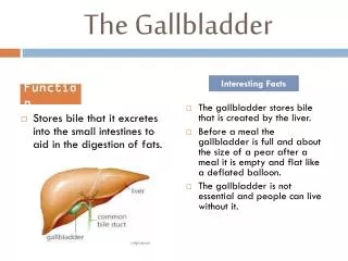
MALIGNANT TUMORS • The incidence of gallbladder carcinoma in the United States is 2.5 cases per 100,000 population • More than 6950 new cases diagnosed per year • Gallbladder carcinoma is the sixth most common carcinoma of the digestive tract • It accounts for 3% to 4% of all gastrointestinal tumorsis • The most common carcinoma of the biliary tree
Epidemiology • Gallbladder carcinoma occurs primarily in the elderly • Three to four times as common in women as in men • The association between gallbladder cancer and gallstones is well established • At least 80% of patients with gallbladder cancer have gallstones • However, gallbladder cancer develops in less than 0.5% of patients with gallstones
Risk factors for gallbladder carcinoma • The risk that gallbladder cancer will develop in patients with gallstones over 20 years is less than 0.5% for the overall population and 1.5% for high-risk groups. The association is probably related to chronic inflammation of the gallbladder; larger stones (>3 cm) are associated with a 10-fold higher risk of cancer than smaller stones • Other risk factors for gallbladder carcinoma are a calcified gallbladder • A long common channel formed by the union of the pancreatic and common bile ducts • The chronic typhoid carrier state
Histological subtypes • Histologically, approximately 80% of gallbladder carcinomas are adenocarcinomas • Histologic subtypes include papillary, nodular, and tubular adenocarcinomas • Papillary tumors, which grow predominantly intraluminally, have a better prognosis than the other subtypes • Less common types of gallbladder carcinoma include squamous cell carcinoma, cystadenocarcinoma, small cell carcinoma, and adenoacanthoma
Metastases • Gallbladder carcinoma spreads via both lymphatic and venous vessels • Because the cholecystic veins drain directly into the liver, gallbladder cancers frequently extend directly into the hepatic parenchyma, usually segments IV and V • Lymphatic spread occurs first to the cystic duct (Calot's) lymph node, then to pericholedochal and hilar nodes, and finally to peripancreatic, duodenal, periportal, celiac, or superior mesenteric nodes • Often the nodal disease of the porta hepatis leads to biliary obstruction and presentation with jaundice • Not surprisingly, presentation with jaundice is associated with a dismal prognosis, even if the patient is otherwise asymptomatic
Clinical presentation • The clinical presentation of gallbladder carcinoma ranges from an incidental finding to a rapidly progressive disease that affords little opportunity for effective treatment • Symptoms and signs associated with gallbladder cancer are commonly nonspecific and include abdominal pain, nausea, fatty food intolerance, anorexia, weight loss, fever, and chills • The most common presenting symptom is right upper quadrant pain, which is present in more than 80% of patients • As the disease advances, the pain often becomes continuous; obstruction of the common bile duct with jaundice may occur in up to 30% of patients • Physical findings in advanced cases include right upper quadrant tenderness, a palpable mass, hepatomegaly, and ascites • Laboratory findings are often unremarkable until obstructive jaundice develops • There are no reliable tumor markers for early detection of this disease
Diagnosis • Abdominal ultrasonography is usually the initial diagnostic procedure • Thickening of the gallbladder wall or a polypoid mass should raise the suspicion of a gallbladder neoplasm • Computed tomography (CT) is more sensitive than ultrasonography, delineates a gallbladder mass with sensitivity and specificity rates of nearly 90% • Is critical for determining resectability, the extent of local disease, and the presence or absence of lymphadenopathy, hepatic metastases, and invasion of the portal vein and hepatic artery by tumor • Magnetic resonance imaging (MRI), specifically magnetic resonance cholangiopancreatography (MRCP), permits noninvasive assessment of the hepatic parenchyma, biliary tree, vasculature, and lymph nodes • Endoscopic ultrasonography is also useful, especially for demonstrating the extent of tumor invasion and lymph node metastases • In patients who present with obstructive jaundice, either endoscopic retrograde cholangiopancreatography (ERCP) or percutaneous transhepatic cholangiography (THC) can identify the level and extent of biliary obstruction
Diagnosis • The tumor is typically detected in one of the following three ways: • (1) as an incidental finding during or after cholecystectomy for suspected benign disease • (2) as a suspected or confirmed neoplasm that appears to be resectable after preoperative evaluation • (3) as an advanced, unresectable intra-abdominal malignancy
American Joint Committee on Cancer Staging of Gallbladder Carcinoma • Primary Tumor (T stage) • T1Tumor invades lamina propria or muscle layer • T1a: Tumor invades lamina propria • T1b: Tumor invades muscle layer • T2Tumor invades perimuscular connective tissue • T3Tumor perforates serosa or directly invades one adjacent organ (extension £2 cm into liver) • T4Tumor extends ≥2 cm into liver and/or into two or more adjacent • Regional Lymph Nodes (N Stage) • N0No regional lymph node metastasis • N1Metastasis in cystic ductal, pericholedochal, and/or hilar lymph nodes • N2Metastasis in peripancreatic, periduodenal, periportal, celiac, and/or superior mesenteric lymph nodes • Distant Metastasis (M Stage) • M0No distant metastasis • M1Distant metastasis
TNM Stage Grouping • Stage I T1 N0 M0 • Stage II T2 N0 M0 • Stage III T3 N0 M0 or • T1-3 N1 M0 • Stage IVAT4 N0-2 M0 • StageIVBT1-4 N0-2 M1
Surgical Resection • If preoperative evaluation suggests gallbladder carcinoma, laparoscopic cholecystectomy should be avoided, because it may worsen the prognosis through incomplete excision of the tumor and spillage of bile • More commonly, carcinoma is not suspected preoperatively, and the cancer is found at the time of laparoscopy for cholelithiasis , in this case, biopsy of the gallbladder mass should be avoided, and conversion to open laparotomy should be performed • If cancer is diagnosed on histologic examination of the gallbladder after laparoscopic resection, further management is based on the stage of the tumor • If reoperation is performed, all trochar sites should be excised in their entirety
Surgical Resection • In patients with stage I gallbladder cancer (TIN0M0), disease is confined to the gallbladder wall, and a simple cholecystectomy is adequate provided that the cystic duct margin is negative for tumor. In most series, simple cholecystectomy is associated with a survival rate of nearly 100% • The management of patients with T2 and T3 lesions is generally accepted to involve extended or radical cholecystectomy, which consists of en bloc resection of the gallbladder and nonanatomic wedge resection of the gallbladder bed (segments IV and V of the liver), with at least a 3- to 4-cm margin of normal liver parenchyma. Regional lymphadenectomy of the choledochal, periportal, hilar, and high pancreatic lymph nodes should be performed • The management of T4 lesions remains controversial. Several series have demonstrated that radical resections for gallbladder cancer can be performed with mortality rates of less than 4%. In these series, at least 50% of patients had advanced T-stage disease (T3 and T4), and 5-year survival rates ranged from 31% to 65%
Palliation • If unresectable local disease is found at the time of exploration, a biliary bypass (hepaticojejunostomy) can be performed to relieve extrahepatic biliary obstruction and the associated pruritus, jaundice, and progressive liver dysfunction • If disseminated disease is found at laparotomy, at laparoscopy, or preoperatively, biliary drainage can be achieved with placement of either a percutaneous or endoscopic stent • Placement of an expandable metal stent can provide permanent internal decompression of biliary obstruction in patients with a life expectancy of only a few months
Chemotherapy and Radiation Therapy • Gallbladder carcinoma is believed to be resistant to most standard chemoradiation regimens in the neoadjuvant (preoperative), adjuvant (postoperative), and palliative settings • The most commonly used chemotherapeutic agent has been 5-fluorouracil (5-FU), with associated response rates ranging from 5% to 30% in most series • In the adjuvant setting, radiation therapy is used to control microscopic residual foci of carcinoma in the tumor bed. Approaches have included standard external-beam radiation therapy, intraoperative external-beam radiation therapy (IORT), and brachytherapy • The benefit of radiation in the palliative setting is modest. The median survival is 6 months after palliative surgery and 2 months after biopsy of the tumor alone without treatment. The addition of palliative radiation therapy increases the median survival to 4 months after biopsy without surgery and to 8 months after palliative surgery
Prognosis • The overall 5-year survival rate for patients with gallbladder carcinoma is less than 5%, with a median survival of less than 6 months • The overall survival rate in series of patients who have undergone resection and whose tumors have been staged according to the AJCC system is nearly 100% for stage I and nearly 50% for node-negative stage II and stage III disease • Although aggressive surgical resection in large centers has offered some improvement in these results, most patients still present with advanced disease that is unlikely to be amenable to cure
BENIGN TUMORS • Benign tumors of the gallbladder manifest most commonly as polyps or polypoid lesions. Polyps can be adenomas, pseudotumors, or hyperplastic inflammatory lesions • Adenomas of the gallbladder are rare and can be sessile or polypoid, they may be premalignant • Cholesterolosis, or pseudotumors, is manifested by yellow spots visible on the surface of the gallbladder mucosa that give the appearance of a “strawberry gallbladder • Inflammatory polyps are composed of a vascular connective tissue stalk with a single layer of columnar epithelial cells • Adenomyomatosis of the gallbladder is characterized by the proliferation of the mucosa and hypertrophy of the underlying muscular layers • The management of a benign gallbladder tumor depends on the size of the lesion. Because polyps larger than 1 cm have the greatest malignant potential, even asymptomatic patients with such polyps should undergo cholecystectomy • Tumors smaller than 1 cm have been shown not to progress to carcinoma; therefore, routine follow-up cholecystectomy is not warranted