Download
1 / 33
360 likes | 551 Views
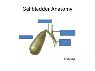
Gallbladder. Rivera, Rivere , Robosa , Rodas , Rodriguez, Rogelio. General Data. 89-year old/ female Chief complaint: severe abdominal pain. . History of the Present Illness: . Past Medical History: .

E N D
Gallbladder Rivera, Rivere, Robosa, Rodas, Rodriguez, Rogelio
General Data • 89-year old/ female • Chief complaint: severe abdominal pain.
Past Medical History: • The patient has diabetes mellitus, hypertension, gastroesophageal reflux disease (GERD), and sick sinus syndrome with a pacemaker • Patient's medications: • metformin, indapamide, pantoprazole, and aspirin
Physical Examination • lethargic but opens her eyes when called; sleepy but arousable and conversant • not oriented to time or place and slips back into sleep when not actively spoken to • T – 36oC BP - 100/67 mm Hg PR - 100 bpm RR - 18 cpm O2 saturation - 96% on room air • Head and Neck - unremarkable • Heart - paced rhythm with a 3/6 holosystolic murmur heard best at the right upper sternal border • Lungs - coarse rales at bilateral bases • Abdomen - distended and tender mostly at the right upper and lower quadrants; ⊕ guarding but ⊖ rebound tenderness • The remaining physical findings are unremarkable
Salient Features • 89 y/o, female • Severe abdominal pain • (+) gastroesophageal reflux disease • (+) diabetes mellitus • (+) hypertension • Intake of metformin, indapamide, pantoprazole, and aspirin • Not oriented to time or place • On PE: • Lethargic but opens her eyes when called; sleepy but arousable and conversant • Distended and tender abdomen mostly at the right upper and lower quadrant; ⊕ guarding but ⊖ rebound tenderness • Lungs: coarse rales at bilateral bases
Missing Information • Onset of symptoms (simultaneous?) • Temperature • Character of pain • Location and radiation of pain • Character of vomitus and its frequency • Precipitating and relieving factors of the symptoms • Presence/absence of a palpable tender mass on RUQ • Bowel sounds • Murphy’s sign
Differential Diagnosis cholangitis
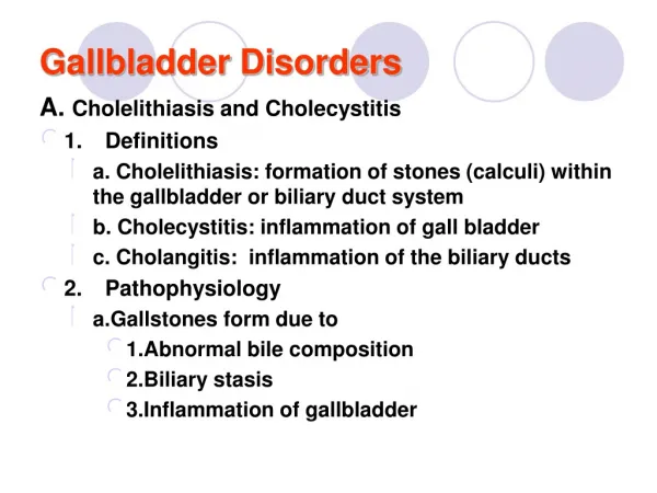
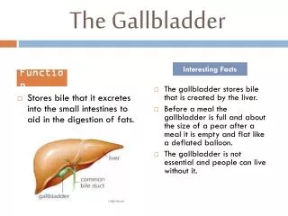
Acute Cholecystitis • Acute cholecystitis occurs as a result of inflammation of the gallbladder wall, usually as a result of obstruction of the cystic duct • In 90% of cases, AC is initiated by the impaction of a calculus in the neck of the GB or in the cystic duct • Pathophysiology: An acute inflammation of the gallbladder caused in most instances by obstruction of the cystic duct, resulting in acute inflammation of the GB wall --the usual cause of the obstruction is a gallstone
Chronic Cholecystitis • Asymptomatic for years, may progress to symptomatic gallbladder disease and may present with complications • Empyema and hydrops • Gangrene and perforation • Fistula formation and gallstone ileus • Limey bile and porcelain gallbladder
Cholangitis • Bacterial infection superimposed on an obstruction of the biliary tree most commonly from a gallstone • Also associated with neoplasms or strictures • Pathophysiology: biliary tract obstruction, elevated intraluminal pressure, and infection of bile
Clinical Impression: Acute Cholecystitis • In elderly patients and in those with diabetes mellitus, acute cholecystitis may have a subtle presentation resulting in a delay in diagnosis • Diabetic patients may have fewer symptoms because of their neuropathy • Incidence of complications is higher in these patients, who also have approximately tenfold the mortality rate compared to that of younger and healthier patients Brunicardi, et al. 2004. Schwartz’s Principles of Surgery, 8th ed. Goldman, Ausiello. 2007. Cecil Medicine, 23rd ed.
Emphysematous Cholecystitis • Presence of air – acute cholecystitis resulting from gall bladder infection with a gas forming organism • pneumoperitoneum
air in gall bladder wall is diagnostic of this disease • Wall thickening • Pericholecystic fluid
Emphysematous Cholecystitis • acute infection of the gallbladder wall caused by gas-forming organisms • Pathogenic factors: • Vascular compromise of the gallbladder • Gallstones • Impaired immune protection • Infection with gas-forming organisms
Diagnosis: Emphysematous Cholecystitis secondary to AcalculousCholecystitis • Vascular compromise to the gallbladder and the presence of gas-forming bacteria, which are more common in patients who have diabetes or are immunocompromised, can cause gangrenous or emphysematous cholecystitis. • Clinically, patients are usually very ill with a high temperature, features of systemic sepsis, and obtundation. • Recommendation: Ultrasound to rule out calculous etiology Goldman, Ausiello. 2007. Cecil Medicine, 23rd ed.
Management • Given that the patient is: • Elderly (89 y/o) • Diabetic (immunosuppressed) • Hemodynamically unstable
Management • Medical • In-hospital stabilization • Surgical • Definitive treatment
Management • STABILIZE: Resuscitate • Repair extracellular volume depletion and electrolyte abnormalities • For analgesia: Meperidine or NSAIDs • less spasm of sphincter of Oddi compared to morphine • Give IV antibiotics (severe case – emphysematous cholecystitis) • Most common organisms likely to be present: E. coli, Klebsiella, Streptococcus • 3rd generation cephalosporins, piperacillin, ampicillinsulbactam, ciprofloxacin + metronidazole • Also for broader coverage since patient is immunosuppressed Fauci, et al. 2008. Harrison’s Principles of Internal Medicine, 17th ed. Brunicardi, et al. 2004. Schwartz’s Principles of Surgery, 8th ed. Goldman, Ausiello. 2007. Cecil Medicine, 23rd ed. Bloom AA. 2008. Emphysematous Cholecystitis: Treatment and Medication. <http://emedicine.medscape.com/article/173885-treatment
Management • Cholecystectomy: Definitive treatment • Interventional radiology • Percutaneouscholecystostomy
Management • Cholecystectomy: Definitive treatment • Emphysematous cholecystitis is an emergency situation treated surgically • According to Schwartz, studies have shown that unless the patient is unfit for surgery, early cholecystectomy should be recommended as it offers the patient a definitive solution in one hospital admission, quicker recovery times, and an earlier return to work. • Since patient is unfit for surgery, • 89 years old – high surgical risk • Patient presented late (3-4 days of illness) Fauci, et al. 2008. Harrison’s Principles of Internal Medicine, 17th ed. Brunicardi, et al. 2004. Schwartz’s Principles of Surgery, 8th ed. Goldman, Ausiello. 2007. Cecil Medicine, 23rd ed. Bloom AA. 2008. Emphysematous Cholecystitis: Treatment and Medication. <http://emedicine.medscape.com/article/173885-treatment
Management • Interventional radiology • Percutaneouscholecystostomy • Objective is to drain out the infected material or pus from the gall bladder in order to delay or obviate the surgical operation. • It is performed under local anesthesia, and it takes approximately 45 minutes to 1½ hours. • A needle is inserted into the gall bladder under ultrasound guidance. The same needle puncture site is serially dilated and finally a larger bore soft catheter is placed into the gall bladder. • The catheter may be either a self-retaining catheter, or it may be sutured in place,andbe connected to a bag. • Clinical improvement in ¾ of patients http://www.pubmedcentral.nih.gov/articlerender.fcgi?artid=1124163
THE CHANGING FACE OF EMPHYSEMATOUS CHOLECYSTITIS K S GILL,MRCP, A H CHAPMAN,FRCR and M J WESTON FRCRDepartment of Radiology, St James’s University Hospital, Beckett Street, Leeds LS9 7TF, UK
OBJECTIVE To describe experiences with cases of emphysematous cholecystitis using newer and more sensitive imaging modalities. SUMMARY BACKGROUND DATA • In the past, the diagnosis has relied on the plain abdominal radiograph (AXR), since there are no clinical features to separate this condition from simple acute cholecystitis. The apparently high mortality and morbidity associated with emphysematous cholecystitis has previously emphasized the importance of emergency cholecystectomy. • We have reviewed eight cases of emphysematous cholecystitispresenting to this hospital over the last 5 years. The diagnosis was made on AXR in only one of these cases. Ultrasound (US) scans were performed in all eight cases, of which five were positive and three negative, due to non visualization of the gall bladder. In the three negative cases, the diagnosis was made on subsequent CT scans.
METHODS A computer search of all radiological reports in our hospital spanning the last 5 years key words ‘‘emphysematous cholecystitis’’ Eight patients had the diagnosis of emphysematous cholecystitis confirmed clinical presentation, investigation, management and progress have been evaluated and correlated with relevant radiology
FINDINGS AXR • 7 of 8 • In 3 cases, reported as normal • 1 case reported as being suspicious of being biliary tract gas on AXR and subsequent US confirmed the presence of emphysematous cholecystitis • In 3 cases, AXR was performed following US or CT and confirmed the diagnosis US • All 8 patients • 5 cases-primary diagnosing modality • 3 cases-failed to diagnose, CT was required
FINDINGS CT • Diagnostic in 3 cases where US failed to make the diagnosis • Established the diagnosis in 4 cases that it was used • Average delay before diagnosis was 2 days from admission • Delay was less (less than 1 day on average) if the diagnosis was made with US • Average delay of 4 days when the diagnosis was made or confirmed by CT because in these cases, CT was only requested subsequent to a non diagnostic US examination
DISCUSSION • Until recently the only means of preoperative diagnosis was the AXR since there are no clinical diagnostic features to separate this condition from simple acute cholecystitis. • Our experience of 8 cases seen in a single hospital over a 5 yr period seems to indicate that this condition is less rare than earlier reports have suggested. The explanation for the discrepancy is considered to be the increasing use of US, and to a lesser extent, CT early in the diagnosis of hepatobiliary disease. CONCLUSION • US and CT are much more sensitive in identifying this condition than the plain AXR. However, US is far from being 100% sensitive. • Whilst CT appears much closer to being 100% sensitive in this small series, it is not a routine investigation in the diagnosis of suspected acute cholecystitis. However, CT is appropriate in cases of failed, or inadequate gall bladder visualization on US. • Consequently, only the severe end of the spectrum was recognized by AXR and this provoked the reports of emphysematous cholecystitis being a rapidly progressing disease requiring early surgical intervention.