Download

1 / 35
570 likes | 1.78k Views
STAINS AND STAINING TECHNIQUES. BY SAMUEL AGUAZIM(MD). Why we should be Stain Bacteria : .

E N D
STAINS AND STAINING TECHNIQUES BY SAMUEL AGUAZIM(MD)
Why we should be Stain Bacteria : • Bacteria have nearly the same refractive index as water, therefore, when they are observed under a microscope they are opaque or nearly invisible to the naked eye. Different types of staining methods are used to make the cells and their internal structures more visible under the light microscope
Staining helps in observation of Bacteria • Staining helps in observation of Bacteria Microscopes are of little use unless the specimens for viewing are prepared properly. Microorganisms must be fixed & stained to increase visibility, accentuate specific morphological features, and preserve them for future use
Stains and Staining : • Stains and Staining Bacteria are slightly negatively charged at pH 7.0 Basic dye stains bacteria Acidic dye stains background Simple stain Aqueous or alcohol solution of single basic dye 5
What is a Stain : • A stain is a substance that adheres to a cell, giving the cell color. The presence of color gives the cells significant contrast so are much more visible. Different stains have different affinities for different organisms, or different parts of organisms They are used to differentiate different types of organisms or to view specific parts of organisms
Staining Reaction • Stains - salts composed of a positive and negative ion, one of which is colored (chromophore) • Basic Dyes - chromophore is the positive ion • dye+Cl- • Acid Dyes - chromophore is the negative ion • Na+dye-
Bacteria are slightly negative, so are attracted to the positive chromophore of the BASIC DYE • Common Basic Dyes • crystal violet • methylene blue • safranin • basic fuchsin
Acid Dyes - used for Negative Staining (background is stained) • Mordant - intensifies the stain or coats a structure to make it thicker and easier to see after it is stained • Example: • Flagella - can not normally be seen, but a mordant can be used to increase the diameter of the flagella before it is stained • Salmonella typhosa
Staining Techniques : • Staining is an auxiliary technique used in microscopy to enhance contrast in the microscopic image. Stains and dyes are frequently used in biology and medicine to highlight structures in biological tissues for viewing, often with the aid of different microscopes
Smearing out of the sample : • Smearing out of the sample Fixation : • Fixation–which may itself consist of several steps–aims to preserve the shape of the cells or tissue involved as much as possible. Sometimes heat fixation is used to kill, adhere, and alter the specimen so it will accept stains Simple staining : • Simple staining simplest, the actual staining process may involve immersing the sample (before or after fixation and mounting) in dye solution, followed by rinsing and observation. The stain can be poured drop by drop on the slide
Simple Bacteria have nearly the same refractive index as water, therefore, when they are observed under a microscope they are opaque or nearly invisible to the naked eye. Different types of staining methods are used to make the cells and their internal structures more visible under the light microscope. Simple stains use one dye that stains the cell wall. The cells are then visible against a light background. Steps: • Place the slide on the staining rack. • Flood the slide with a basic stain: either crystal violet (1 min.), Safranin (2 min.), or Methylene blue (2 min.). • Wash the stain off the slide with deionized water. • Blot the slide with bibulous paper.
Simple staining (cont..) : • Simple staining (cont..) Dilute Carbolfuchsin:- Made by diluting Z-N stain with 10- 15 times its volume of water- Stain for 20-25 seconds, wash with water Use: To demonstrate the morphology of Vibrio cholera Polychrome methylene blue: Use: M’Fadyean’s reaction - B. anthracis
Simple Stains : • Simple Stains Bacterial arrangement : • Bacterial arrangement Clusters (group). Chains. Pairs (diploids). No special arrangement. • Simple Staining Easier to Perform But has Limitations : • Simple Staining Easier to Perform But has Limitations Simple easy to use; single staining agent used; using basic and acid dyes. Features of dyes: give coloring of microorganisms; bind specifically to various cell structures
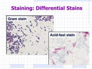
Differential Stains : • Differential Stains use two or more stains and allow the cells to be categorized into various groups or types. Both techniques allow the observation of cell morphology, or shape, but differential staining usually provides more information about the characteristics of the cell wall (Thickness). Most Common • Gram Stain • Acid-Fast Stain
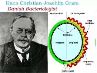
GRAM STAIN • 1884 Hans Christian Gram • most important stain used in Bacteriology • Divides all Bacteria into 2 groups: • Gram (+) • Gram (-)
GRAM STAIN 1. Crystal violet
GRAM STAIN • 2. Grams Iodine (mordant)
GRAM STAIN • 3. Alcohol
GRAM STAIN • 4. Safranin (Counterstain)
RESULT • Gram (+) BLUE • Gram (-) RED • Difference - due to structure of cell wall • Gram (+) Thick cell wall • Gram (-) Thin cell wall
Identification of a Bacteria Unknown • 1. Gram Reaction • 2. Morphology
Acid - Fast Stain • Differential Stain - divides bacteria into 2 groups • Acid - Fast • Non Acid - Fast • Used to identify organisms in the Genera Mycobacterium(high lipid and wax content in cell wall)
Acid - Fast Stain • 1. Carbolfuchsin(Red) • 2. Acid Alcohol • 3. Counterstain with Methylene Blue • Acid - Fast Cells Red • Non Acid - Fast Blue
Special Stains Capsule Stain Klebsiellapneumoniae
Flagella Stain Spirillumvolutans
Endospore Stain Bacillus cereus
Bacterial Cell Shapes • Bacteria can have several different shapes, but the primary shapes we will be observing are: • Spherical or round cells – cocci (plural) or coccus (singular) • Rod shaped – bacilli (plural) or bacillus (singular) • Spiral shaped – spirilla • Some bacteria have characteristic clustering or arrangements, usually due to how the cells divide and whether they remain attached together when they divide. • Diplococci – divide in one plane and remain attached together after cell division. • Streptococci – divide in one plane and form long chains of attached cells. • Staphylococci – divide in many planes and remain attached together forming a “grape-like cluster”
Gram-positive : • Gram-positive bacteria are those that are stained dark blue or violet by Gram staining. This is in contrast to Gram-negative bacteria, which cannot retain the crystal violet stain, instead taking up the counter stain (safranin or fuchsine) and appearing red or pink. Gram-positive organisms are able to retain the crystal violet stain because of the high amount of peptidoglycan in the cell wall. Gram-positive cell walls typically lack the outer membrane found in Gram-negative bacteria.
GRAM-POSITIVE BACTERIA : • GRAM-POSITIVE BACTERIA are characterized by having as part of their cell wall structure peptidoglycan as well as polysaccharides and/or teichoic acids. The peptidoglycans which are sometimes also called murein are heteropolymers of glycan strands, which are cross-linked through short peptides.
What are Gram Negative Bacteria • Bacteria Gram-negative bacteria are those bacteria that do not retain crystal violet dye in the Gram staining protocol. In a Gram stain test, a counter stain (commonly safranin) is added after the crystal violet, coloring all Gram-negative bacteria with a red or pink color. The test itself is useful in classifying two distinct types of bacteria based on the structural differences of their cell walls. On the other hand, Gram-positive bacteria will retain the crystal violet dye when washed in a decolorizing solution
Gram negative bacteria • Gram negative bacteria On most Gram-stained preparations, Gram-negative organisms will appear red or pink because they are counterstained. Due to presence of higher lipid content, after alcohol-treatment, the porosity of the cell wall increases, hence the CVI complex (Crystal violet -Iodine) can pass through. Thus, the primary stain is not retained
THANK YOU • THANK YOU!!!!!!!!!!!!!!!!!!!!!!!!!!!!!!!!!!!!!!!!