Download

1 / 25
1.12k likes | 5.01k Views
Differential Staining Techniques. By Elif TEKİN. Types of Staining . Different types of staining methods are used to make the cells and their internal structures more visible under the light microscope. Simple stain ing

E N D
Differential Staining Techniques By Elif TEKİN
Types of Staining • Different types of staining methods are used to make the cells and their internal structures more visible under the light microscope. • Simple staining colors all cells of specimen in one type. Only one stain isused and the aim is creating contrast for examination of cell morphology • Differential staining colors cells by showing their different composition or structures.
Differential Staining • use two or more stains and allow the cells to be categorized into various groups or types. • at least2 types of stains are used the first stain is called the primary stain and the second stain is called counterstain. • are used for identification or structure examination of bacteria in addition to general morphological examination
Differential Staining • Gram Staining • Endospore Staining • Capsule Staining
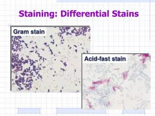
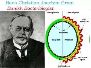
Gram Staining • Gram staining is a differential staining technique that provides aneasy differentiation of bacteria into one of two groups. Gram positive and Gram negative • Basic classification of bacteria is based on the cell wall structure. • The amount of peptidoglycan, the characteristic compound found in all true bacterial cell walls, is among one of the differences between the Gram Positive and Gram Negative cell walls.
Gram Staining Gram-positive cell walls Gram-negative cellwalls • Thick peptidoglycan • 90% peptidoglycan • Teichoicacids • Not many polysaccharides • Thin peptidoglycan • 5-10% peptidoglycan • No teichoicacids • Outer membrane has lipids, polysaccharides
Gram Staining There are 4 conditions to be followed for a valid Gram staining procedure: Young cultures - must be young within 18-24hrs old (older cultures lose their Gram staining properties due to changes in the CWs as the cells get older) Thin smear thicker or uneven smears will result in uneven staining and decolorization Fresh reagents - of proper strength Control cultures - for a known GP bacterium and GN culture (S.aureus & E.coli)
Remember! Too thin smears are obtained usually with NB cultures. Few cells on the slide to observe and it would be difficult to find them. Also you may lose them easily during staining. It happens when: - The culture is taken from NB or NA that is not dense enough - The culture is taken from upper part of NB tube without shaking - Heat fixation performed less than enoughcells are washed away. Too thick smears are obtained usually with NA cultures. Too many bacterial cells on the slide. This prevents proper light penetration so lessening visibility. Also it would be difficult to discriminate individual cells and determine their sizes, shapes and structures. It happens when: - More than enough cells are taken from colonies of NA. (less than a single colony is OK) - Culture is taken from bottom part of NB tube without shaking .
Remember! • Distorted cells can be observed if heat fixation is done more than enough (3-4 times are OK). So the cells are seen different than their original sizes and shapes. If the smear is heated much more, all the cells coagulates and form a mass of molecules not single cells
Gram Staining Procedure Preapare smears then; 1. Apply few drops of crystal violet to cover the smearfor 1 min 2. Pour off excess dye and Rinse with dH2O 3. Then cover the smear with Gram’s Iodineand leave it 1,5min. 4. Rinse with dH2O. 5. Wash with 95% Ethanol (decolorizing agent) until fluid flows off the smear is colorless 6. Rinse immediatelywith dH2O. 7. Apply safranin to smear and wait 1 min. 8. Wash with dH2O and Air-dry Observe under100x objective and Notecolors, shapes,sizes and staining properties of cells.
Gram Staining Pseudomonas Neisseria Streptococcus
Gram Staining Gram (-) cell are always seen as Gram (-) but Gram (+) cells can beseen as Gram (-) due to someexperimental errors. This unexpected result is called Gramvariablereaction. Reasons for Gram-variable reaction: 1. Age of the culture stained with Gram staining should be smaller than 24 h. It should benot damaged by chemicals or not treated by antibiotics. 2. Too thick smears may cause improper decolorization. So smear thickness should be proper. 3. Too much heat fixation causes cells to rupture. In this way Gram (+) cells may loseCV-I and can take Safranin color.
Gram Staining 4. Fresh chemicals should be used in proper concentrations. Applications of low amountor concentrations of Crystal violet, iodine solution or safranin may cause Gram (+) cells stained as Gram (-). 5. Nature, concentration and amount of decolorizing agent is very important. If youapply too much, you cause Gram (+) cells to lose CV-I complex and appear in pink..Also prolonged washing between each step may cause over-decolorization
Endospore Staining • Spore: structure that can survive for long periods of time in a metabolically inert state • Produced when environmental conditions are poor • Do not stain well with simple stains harsher protocol required (i.e. heat)
Endospore Staing (Schaffer and Fulton staining method) • Malachite Green is used to stain the sporeas primary stain. • Steam is applied to enhance penetration of dye to relatively impermeablespore coat. It has low affinity to ordinary dyes. • After that slides are cooled to trap stain inside,resisting decolorisation. Rinsing with water decolorizes MG from vegetative cells. • Safranin isused as counterstain to stain the colorless vegetative part. • At the end of the staining the sporesappear in green and vegetative parts appear in red.
Endospore Staining Prepare smear! 1. Put the slide to tripod places in staining rack. 2. Cover the smear with Malachite Green and wait1 min. 3. Pass flame under slide quickly for manytimes. It will take about 3-5 min. Do not drythe smear Pay attention to not to boil the smear. Heat ituntil you observe vapor after removal of flame 4. Cool the slide and Wash with dH2O. 5. Apply Safranine and wait for 1 min. 6. Wash with dH2O. Then perform blotting. Observe the specimen with 100x objective. Try to differentiate spores and vegetative parts.
Capsule Staining Capsule is a polysaccharide-containing material lying outside of the bacterial cell. It is a kindof glycocalyx secreted by cytoplasm. The traditional capsule is the one forming a kind of tight matrix excluding large particles. Its functions are related mostly with pathogenecity: • It aids attachment of certain pathogenic microorganisms to host. • Engulfment of pathogens by phagocytic cells of immune system becomes difficult with presence of capsule. • It has role in preventing dessication by binding some water.
Capsule Staining Negative Staining performed in order to observe capsule lying outside the cell. Since mostcapsules are largely water soluble and nonionic, they do not bind ordinary stains. Also heatapplication of ordinary smear preparation damages the capsule.
Capsule Staining • Indian ink is used to color the background. Its particles are too largeto pass from capsule so it is excluded. • Safranin is used to color cells. So thecolorless uniform zone between pink cell and dark background is the capsule. Not all clearzones are capsules. Shrinkage of cells, withdrawal of ink and cracks cause someclear zones to form. Capsuleis seen as a uniform clear zone.
Capsule Staining Protocol 1. Place 1 drop of Indian ink near one edge of a clean slide. 2. Aseptically get a loop of culture given toyou and mix it with the ink drop on slide. 3. Use a second slide as spreader andspread the mixture first by touching theshort edge of spreader to the mixture. Let the mixture run across the edge andthen spread the liquid across the lengthof the slide by holding the spreadertilted. 4. Let it air dry completely,no heat fixation and no washing, Heat detoriates capsule 5. Cover the smear with Safranin and wait for 1 min. 6. Rinse with dH2O. 7. Perform blotting and air dry if necessary. 8. Observe with 100x objective
CAPSULE STAINING 1. Place 1 drop of Indian ink near one edge of a slide. 2. Aseptically get a loop of culture given toyou and mix it with the ink drop on slide. 3. Use a second slide as spreader. 4. Let it air dry completely,no heat fixation and no washing, Heat detoriates capsule 5. Cover the smear with Safranin and wait for 1 min. 6. Rinse with dH2O. 7. Perform blotting and air dry if necessary. ENDOSPORE STAINING 1. Put the slide to tripod places in staining rack. 2. Cover the smear with Malachite Green and wait1 min. 3. Pass flame under slide quickly for manytimes. It will take about 3-5 min. Do not drythe smear Pay attention to not to boil the smear. Heat ituntil you observe vapor after removal of flame 4. Cool the slide and Wash with dH2O. 5. Apply Safranine and wait for 1 min. 6. Wash with dH2O. Air dry GRAM STAINING 1.Apply few drops of crystal violet to cover the smearfor 1 min 2. Pour off excess dye and Rinse with dH2O 3. Then cover the smear with Gram’s Iodineand leave it 1,5 min. 4. Rinse with dH2O. 5. Wash with 95% Ethanol (decolorizing agent) until fluid flows off the smear is colorless 6. Rinse immediatelywith dH2O. 7. Apply safranin to smear and wait 1 min. 8. Wash with dH2O and Air-dry
Group 1,3 Gram staining B. cereus, E.coli Endospore staining B. subtilis Capsule Staining K. pneumonia • Group2 Gram staining B. cereus(old), E.coli Endospore staining B. subtilis Capsule Staining K. pneumonia • Group 4 Gram staining Serratia marcescens, E.coli Endospore staining B. subtilis Capsule Staining K. pneumoniae • Group 5 Gram staining K. pneumoniae, E.coli Endospore staining B. subtilis Capsule Staining K. pneumonia