Download
1 / 17
180 likes | 213 Views
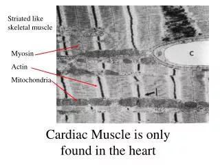
Biology 102 Laboratory 2 Human Heart Anatomy Steer Heart Cardiac Muscle Histology. Objectives for today’s lab. Lab Exam Objectives : After completion of this lab, you should be able to:

E N D
Biology 102 Laboratory 2 Human Heart AnatomySteer Heart Cardiac Muscle Histology
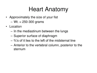
Objectives for today’s lab • Lab Exam Objectives: After completion of this lab, you should be able to: • Identify the anatomical structures of the human heart listed below using heart models, or photographs of models or hearts. (American Heart Model is best) • Identify the anatomical structures of the steer heart listed below using heart models, or photographs of models or hearts.
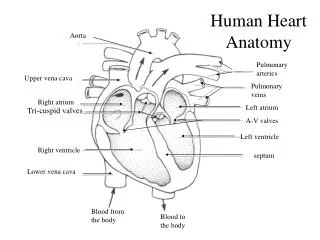
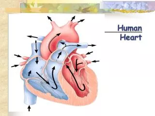
Chambers of Heart/Blood Flow Figure from: Martini, Anatomy & Physiology, Prentice Hall, 2001
Coverings of Heart Figure from: Martini, Anatomy & Physiology, Prentice Hall, 2001 Fibrous pericardium = pericardial sac (outermost layer)
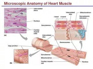
Wall of Heart Figure from: Hole’s Human A&P, 12th edition, 2010 • Three layers • endocardium • forms protective inner lining • membrane of epithelial and connective tissues • myocardium • cardiac muscle • contracts to pump blood • epicardium • serous membrane (visceral pericardium) • protective covering • contains capillaries and nerve fibers Know all the layers depicted in the diagram, and know their correct order.
Heart Valves Pulmonary and Aortic Valve Tricuspid Valve Figures from: Hole’s Human A&P, 12th edition, 2010
Blood Supply to Heart (Coronary circulation) Anastamoses = connections between 2 or more branches of arteries that supply the same region with blood. Give rise to collateral circulation (LAD) Blood flow through coronary arteries takes place mainly during relaxation of the ventricles (ventricular diastole) Figure from: Martini, Anatomy & Physiology, Prentice Hall, 2001
Blood Supply to Heart Cardiac veins join at an enlargement called the coronary sinus that drains into the right atrium Figure from: Martini, Anatomy & Physiology, Prentice Hall, 2001
Brachiocephalic artery Aorta Superior vena cava Pulmonary trunk Right atrium Left atrium Right ventricle Left ventricle Apex of the heart From: Marieb and Mitchell , 10th edition, Pearson
Aortic semilunar valve Aorta Right atrium Left atrium Tricuspid valve Bicuspid valve Myocardium Chordae tendineae Right ventricle Trabeculae carneae Interventricular septum Left ventricle From: Marieb and Mitchell , 10th edition, Pearson
Aorta Left atrium Aortic semilunar valve Bicuspid valve Tricuspid valve Chordae tendineae Right ventricle Left ventricle Interventricular septum Papillary muscles From: Marieb and Mitchell , 10th edition, Pearson
Calf Heart From: http://www.nietvergeten.com/jeroen_advocaat/englisch/medical_illustration.htm#20
Things you should do in lab today • Identify the structures listed on your Laboratory Guide • Human heart models (I suggest you become familiar with the human heart before beginning dissection) • Steer heart
Next Lab… • Blood Vessel Anatomy • See Exercise 32 in Marieb’s Lab Manual • Be sure to review what you did today and complete Review Sheet for Exercise 30 in your lab manual. • You will be combining blood flow in the heart with what you’re learning about blood vessels.