Download
1 / 33
330 likes | 332 Views
Biology 102 Laboratory 3 Arteries I Human/Cat Gross Anatomy Histology. Objectives for Lab 3. Identify the arteries above the diaphragm and in the upper limb listed on your Lab Handout in a human model or photograph of a human.

E N D
Biology 102 Laboratory 3 Arteries I Human/Cat Gross AnatomyHistology
Objectives for Lab 3 • Identify the arteries above the diaphragm and in the upper limb listed on your Lab Handout in a human model or photograph of a human. • Identify the arteries above the diaphragm and in the upper limb listed on your Lab Handout on a dissected cat or photograph of a cat. • Describe the layers/tunics and histological characteristics of arteries. • Be able to trace the path of a drop of blood through the arteries you learned today.
Systemic Circuit • Composed of vessels that lead from the heart to all body parts (except the lungs) and back to the heart • Includes the aorta and its branches (know!) • Includes the system of veins that return blood to the right atrium • Includes the coronary circulation
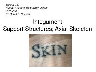
Comparison of Walls of Arteries and Veins Smooth muscle of the tunica media in both arteries and veins is innervated by the sympathetic nervous system. Figure from: Hole’s Human A&P, 12th edition, 2010
Major Vessels of Arterial System • Arteries carry blood AWAY from heart. • Same ‘tube’ is named something different depending on the anatomical landmark it’s near. Figure from: Martini, Anatomy & Physiology, Prentice Hall, 2001
Major Blood Vessels Associated With Heart Figure from: Hole’s Human A&P, 12th edition, 2010 You should know all the vessels and structures on this slide Remember your ABCs: Aortic arch, Brachiocephalic, Carotid, subclavian
Arteries to Neck, Head, and Brain Figure from: Hole’s Human A&P, 12th edition, 2010 * * * * * *
Arteries to Shoulder and Upper Limb * * * = pulse points * * * Figure from: Hole’s Human A&P, 12th edition, 2010
Cat Blood Vessel Dissection Schedule • Blood vessel labs • Dissection 1(today - arteries above diaphragm and upper limb) • Preparing the cat (see page 721 in Marieb) • Cardiovascular (pp. 725-728 in Marieb) • Dissection 2 (next lab - arteries below diaphragm and lower limbs) • Cardiovascular (pp. 725-728 in Marieb) • Dissection 3 (two weeks from today – all veins) • Cardiovascular (pp. 729-732 in Marieb)
Objectives for Today’s Cat Dissection • Begin dissecting the preserved cat to identify arteries above the diaphragm and in the upper limb. • Begin to identify the arteries listed on the Cat Artery Checklist for Lab 3 in your Laboratory Guide • DON’T RUSH YOUR DISSECTION!!!
Precautions for Cat Dissection • You must wear gloves and goggles for this lab! • ABSOLUTELY NO FOOD OR DRINK • Be EXTREMELY careful when using the scalpels • Keep your index finger on the top of the blade • Be careful not to inadvertently cut structures • Use a blunt probe, or your gloved fingers to dissect away tissue. Use the scalpel ONLY for initial cutting. Proceed S-L-O-W-L-Y. • Discard scraps/liquid into the appropriate container • Gloves, paper towels, plastic bags -> regular trash
Getting started with Cat Dissection… • Work in groups of three or four • Get some paper towels and keep them at your lab table • Get your instruments and dissecting tray (large trays in the front of the lab) ready • Get a cat and cut open the sealed bag but don’t discard the fluid; you’ll be using this cat all semester and replacing the fluid each time. • Let the cat out of the bag!
Getting started with Cat Dissection… • See page 721 in Marieb’s Lab Manual • This will prepare the cat for the rest of the dissections • Try to identify the major organs you see before you begin dissecting any further • Look at the preserved, dissected cat (front of lab) to get an idea of what you’ll see for each system
Getting started with Cat Dissection… • Once the initial cuts have been made according to the instructions in your Lab Manual, you are ready to continue with artery identification • Note that the cats have been dual injected with dyes (red = arteries, blue = veins) • Note the differences in the anatomy of the cat (vs. the human) in the blood vessels around the heart (see your Laboratory Handout)
Cardiovascular system • Open the rib cage by cutting just to one side of the sternum with scissors then spread the ribs • Examine the position of the thoracic organs, i.e., heart and lungs and the membranes surrounding them • Gently begin to remove the fatty/glandular tissue from the surface of the heart to expose the vessels around the heart (see preserved cat) • Work S-L-O-W-L-Y to dissect away the CT from the vessels. Use a blunt probe.
Initial Cuts to be made to prepare for the Cat for further dissection
Artery Dissection • You should be able to locate the arteries above the diaphragm and in the upper limb that are listed in your Lab Guide • Notice the pulmonary vessels and try to trace their path to their connections to the lungs
Right common carotid artery Left common carotid artery Right subclavian artery Left subclavian artery Brachiocephalic artery Aorta Heart (within pericardium)
Left external maxillary artery Left lingual artery Left external carotid artery Superior thyroid artery Left internal carotid artery Left common carotid artery
Radial vein Radial artery Brachial artery Ulnar artery Brachial vein
Left thyrocervical artery Left axillary artery Left brachial artery Left vertebral artery Left subclavian artery
Right external jugular vein Left common carotid artery Right common carotid artery Right subclavian vein Left external jugular vein Left subclavian vein Right brachiocephalic vein Right subclavian artery Left brachiocephalic vein Left subclavian artery Superior vena cava Brachiocephalic artery Aorta Right atrium of heart Left ventricle of heart Right ventricle of heart
Right common carotid artery Left common carotid artery Left subclavian artery Right subclavian artery Brachiocephalic artery Aorta
Right common carotid artery Left common carotid artery Right subclavian artery Left subclavian artery Brachiocephalic artery Lungs Descending aorta Ascending aorta Trachea Aortic arch *Note the differences from the human
Brachiocephalic artery Left subclavian artery Aortic arch Ascending aorta Pulmonary trunk Right ventricle Left atrium Descending aorta Left ventricle Esophagus Diaphragm
Right thyrocervical artery Left thyrocervical artery Right brachial artery Left axillary artery Right axillary artery Right vertebral artery Left vertebral artery Right subclavian artery Left internal mammary artery Right thoracodorsal artery Left subclavian artery Right internal mammary artery
Left common carotid artery Right common carotid artery Right subclavian artery Left subclavian artery Brachiocephalic artery Aortic arch Ascending aorta Heart Descending aorta
Brachiocephalic artery Left subclavian artery Aortic arch Ascending aorta Descending aorta (thoracic aorta) Pulmonary trunk Intercostal arteries Heart
Aorta Right atrium Pulmonary trunk Right ventricle Left atrium Coronary vessels Left ventricle Apex of the heart
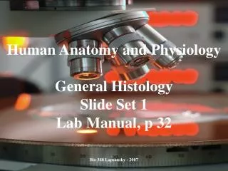
Things you should do in lab today • Examine the artery slide under the microscope • Name/identify the layers of the artery • Describe how each layer (tunic) is constructed • Refer to Marieb’s Lab Manual Figure 32.1 left side • Perform cat dissection of arteries listed for Lab 3 (above diaphragm and upper limb). See Marieb’s Lab manual pp. 721 and 725-728 for instructions. • Review human arteries listed for Lab 3 (above diaphragm and upper limb) • Complete Lab 3 Assignment on a separate sheet of paper, put your name on it, and hand it in today.
To be handed in… • Lab 3 Assignment (Figures 32.2, 32.3, and 32.4) • Exercise 1 - trace the path of a drop of blood from just after it leaves the heart until it reaches left side of the brain. • Exercise 2 - trace the path of a drop of blood from just after it leaves the heart until it reaches right wrist.
Next Lab… • We will continue with arteries in the next lab: those below the diaphragm and serving the lower limb • Good idea to look ahead at the arteries we’ll be covering.