Download
1 / 27
340 likes | 446 Views
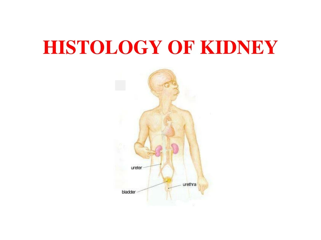
HISTOLOGY OF KIDNEY. MACROSCOPIC STRUCTURE OF KIDNEY. GENERAL ORGANISATION OF KIDNEY. Kidney consists of following parts : 1.Cortex 2.Medulla 3.Major and minor calyces 4.Renal pelvis. Secreting part. Collecting Part. Collecting tubules Ducts of Bellini. Nephrons.

E N D
GENERAL ORGANISATION OF KIDNEY Kidney consists of following parts: 1.Cortex 2.Medulla 3.Major and minor calyces 4.Renal pelvis
Secreting part Collecting Part Collecting tubules Ducts of Bellini Nephrons The kidney is composed of numerous closely packed uriniferoustubules. Uriniferous tubules
CORTEX MEDULLA
CORTEX: • Darker outer portion • Contains renal corpuscles, proximal and distal convoluted tubules and medullary rays • Also contains interlobular arteries and veins
Proximal convoluted tubule: • Lined by simple cuboidal epithelium • Shows prominent brush border • Cytoplasm takes intense eosinophilic staining • Small or uneven lumen
Distal convoluted tubule: • Lined by simple cuboidal epithelium • No brush border • Clear distinct lumen • Less intensely stains • Less in number
Collecting tubules: • Lined by cuboidal epithelium • Becomes progressively taller • No brush border • Collecting ducts: • Formed by the fusion of collecting tubules • Lined by tall columnar epithelium • Pale stained cytoplasm • No brush border
Medulla : • Contains the segments of the loops of Henle, straight portions of tubules, vasa recta and collecting ducts • Thin segments of Henle lined by simple squamous epithelium • Straight portions of tubules are lined by simple cuboidal epithelium • Collecting ducts (ducts of Bellini/papillary ducts) are lined by simple columnar epithelium
CORTEX MEDULLA
Juxtaglomerular apparatus • Macula densa • Lacis cells (cells of Polkissen) • Juxtaglomerular cells
Functions of J.G.Apparatus • Secretion of hormone – renin – increases blood pressure. 2. Stimulates the synthesis and release of aldosterone. 3. Liberates erythropoietic factor which helps maturation of erythrocytes.
Kidney- cortex magnified Capsule Distal convoluted tubule (DCT) Glomerulus Proximal convoluted tubule (PCT) Bowman’s capsule
Kidney- cortex magnified Glomerulus Collecting duct DCT PCT Bowman’s capsule
Muscular tube • Mucosa: transitional epithelium • Shows numerous folds • Wide lamina propria • Muscularis layer: arranged in two layers – inner longitudinal and outer circular • Outer coat: Adventitia – loose connective tissue with blood vessels, nerves and lymphatics URETER
Ureter Circular muscle layer Longitudinal muscle layer Epithelium Adventitia
Ureter Adventitia Blood vessel Transitionalepithelium Lamina propria Lumen Circular muscle layer Longitudinal muscle layer
Identification points • Kidney: • Proximal convoluted tubule is lined by cuboidal cells with microvilli (brush border) and lumen is small. • Distal convoluted tubules are lined by cuboidal epithelium and larger lumen. • Henle’s loop lined by squamous epithelium. • In cortex, presence of glomerulus. • Medulla has collecting ducts lined by columnar cells.
Identification points • Ureter • Mucosa is lined by transitional epithelium. • Inner circular and outer longitudinal layer of smooth muscles can be seen. • Lumen is star shaped. • Outermost layer is adventitia.