Download
1 / 25
360 likes | 958 Views
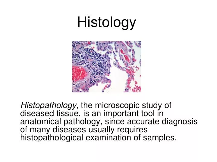
Histology. Histopathology, the microscopic study of diseased tissue, is an important tool in anatomical pathology, since accurate diagnosis of many diseases usually requires histopathological examination of samples. Samples fix in 10% NBF. 48hrs -72 hrs -days. Histology Processing.

E N D
Histology Histopathology, the microscopic study of diseased tissue, is an important tool in anatomical pathology, since accurate diagnosis of many diseases usually requires histopathological examination of samples.
Samples fix in 10% NBF. 48hrs -72 hrs -days Histology Processing
Paraffin infiltration and embedding into blocks. Cuts thin sections on slides (3-5um thick).
Stain tissue sections • Hematoxylin and eosin stain (H & E stain) • structures staining blue are called basophilic because of their affinity for the basic dye (hematoxylin) • structures staining pink are called acidophilic because of their affinity for the acid dye (eosin),
Liver • Hepatocytes – functional liver cells • Hepatic Sinusoids – small blood vessel within liver tissue • Hepatic Portal Vein – brings blood into the liver • Central Vein – takes blood out of the liver • Bile Duct – storage and drainage of bile • Arteriole – delivers blood from artery to capillaries • Glycogen Vacuole – used for glycogen storage
Hepatic sinusoid Hepatocyte Glycogen vacuole Liver 1000X
400x Liver Hepatic sinusoids Erythrocytes visible within the central vein
400X Liver Arteriole
Liver Central vein RBC Arteriole
Liver Bile Duct
Liver Bile Duct
Liver White Blood Cells
Head Kidney • Interrenal gland – Is a cortisol producing tissue.. • Chromaffin cell – neuroendocrine cells that catalyze and secrete epinephrine, norepinephrine, and other hormones • Postcardinal vein – Blood transport • Hematopoietic tissue – where new blood cells are formed
400x A B C Head Kidney • Interrenal Gland • Chromaffin Cell • Postcardinal vein
Head Kidney Cardinal Vein Chromaffin cells Interrenal cells
Trunk Kidney • Nephron – a renal tubule that removes waste and helps maintain homeostasis • Renal corpuscle • Glomerulus – Pressure filters blood plasma into Bowman’s capsule • Basement membrane – glomerulus membrane that does the blood filtering • Bowman’s capsule – collects filtered blood plasma and connects with proximal tubule • Proximal tubule – selectively transports nutrients (amino acids, glucose, salt, ect.) back into the blood • Distal tubule – tubular secretion of molecules to balance pH Melanocytes – pigment cells • Collecting duct and/or Mesonephric duct – collects urea from multiple nephrons and controls futher water reabsorption • Connective tissue – fibres surrounding ducts
Trunk Kidney Distal tubules Proximal tubules Renal corpuscle
Trunk Kidney Basement membrane Bowmans capsule Glomerulus
400x C A B • Mesonephric Duct • Melanocytes • Connective tissue Trunk Kidney
Gills • Filament – threadlike structure forming respiratory surface • Lamella – fingerlike projections of the filament • Goblet (or mucous) cell – secret mucus • Chloride cell – acid/base regulation by Cl- and HCO3- excretion • Cells surface area becomes enhanced during alkalosis and decreases during acidosis • Pillar (or pilaster) cell – lend support for lamelle • Gill filament cartilage – center of filament for structural support
Gill Lamella • Filament cartilage • Pillar cell • Chloride cell C B A Gill filaments and lamellae 40x
Gill Epithelialcells Goblet cells
Gill Chloride cell Pillarcells Erythrocytes
Laboratory Steps • Use proper microscope handling procedures (distributed in Lab 2) • Examine histology slides of liver, kidney, and gills • Examine additional histology slides of other organs