Download

1 / 26
340 likes | 1.2k Views
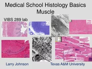
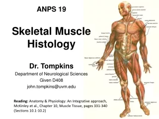
Histology of Muscle. Skeletal Muscle Tissue. Muscle Histology. Elongated cylindrical cells = muscle fibers Plasma membrane = sarcolemma Transverse (T) tubules tunnel from surface to center of each fiber Multiple nuclei lie near surface of cell Cytoplasm = sarcoplasm. Muscle Histology.

E N D
Muscle Histology • Elongated cylindrical cells = muscle fibers • Plasma membrane = sarcolemma • Transverse (T) tubules tunnel from surface to center of each fiber • Multiple nuclei lie near surface of cell • Cytoplasm = sarcoplasm
Muscle Histology • Throughout sarcoplasm is sarcoplasmic reticulum • Stores calcium ions • Sarcoplasm contains myoglobin • Red pigmented protein related to Hemoglobin that carries oxygen • Along entire length are myofibrils • Myofibrils made of protein filaments • Come in thick and thin filaments
The Sarcomere • Filaments overlap in repeating patterns • Unit structure is called sarcomere • Separated by Z discs • Darker area = A band associated with thick filaments • H zone has no thin filaments • I band has thin filaments no thick filaments
Functional Anatomy • Thick filament (myosin) has moveable heads (like “heads” of golf clubs) • Thin filaments (actin) are anchored to Z discs • Contain myosin binding sites for myosin head • Also contain tropomyosin & troponin • Tropomyosin blocks myosin binding site when muscle is at rest
Sliding Filament Mechanism • During contraction myosin heads bind actin sites • Myosins pull and slide actin molecules (and Z discs) toward H zone • I bands and H zones become more narrow • Sliding generates force and shortens sarcomeres and thus fibers
Neuromuscular Interaction • Nerve signal triggers muscle action potential • Delivered by motor neuron • One neuron can trigger 1 or more fibers at the same time • Neuron plus triggered fibers = motor unit
Neuromuscular Interaction • Neuronal ending to muscle fiber = neuromuscular junction (NMJ) • Synaptic end bulbs (at neuron terminal) • Release neurotransmitter • Muscular area = Motor end plate • Between is synaptic cleft
Axon collateral of somatic motor neuron Axon terminal Nerve impulse Synaptic vesicle containing acetylcholine (ACh) Sarcolemma Axon terminal Synaptic end bulb Motor end plate Synaptic end bulb Neuromuscular junction (NMJ) Synaptic cleft (space) Sarcolemma Myofibril (b) Enlarged view of the neuromuscular junction (a) Neuromuscular junction 1 ACh is released from synaptic vesicle 1 Synaptic end bulb Synaptic cleft (space) Motor end plate 2 2 ACh binds to Ach receptor Na+ Junctional fold 3 Muscle action potential is produced (c) Binding of acetylcholine to ACh receptors in the motor end plate
Action at NMJ • Release of acetylcholine (ACh) • Diffuses across cleft • Activation of ACh receptors • Generation of Muscle Action Potential • Repeats with each neuronal action potential • Breakdown of ACh
Contraction Trigger • Muscle action potential →Ca2+ release from Sacroplasmic Reticulum (SR) • Ca2+ binds to troponin→ • Moves tropomyosin off actin sites → • Myosin binds & starts cycle
Contraction Cycle • Myosin binds to actin & releases phosphate group (forming crossbridges) • Crossbridge swivels releasing ADP and shortening sarcomere (power stroke) • ATP binds to Myosin → release of myosin from actin • ATP broken down to ADP & Pi→activates myosin head to bind and start again • Repeats as long as Ca2+ concentration is high
Relaxation • Breakdown of ACh to stop muscle action potentials • Ca2+ ions transported back into SR lowering concentration → • This takes ATP • Tropomyosin covers actin binding sites
1 Nerve impulse arrives at axon terminal of motor neuron and triggers release of acetylcholine (ACh). Muscle action potential Nerve impulse 2 Transverse tubule ACh diffuses across synaptic cleft, binds to its receptors in the motor end plate, and triggers a muscle action potential (AP). 4 Muscle AP travelling along transverse tubule opens Ca2+ release channels in the sarcoplasmic reticulum (SR) membrane, which allows calcium ions to flood into the sarcoplasm. ACh receptor 3 Acetylcholinesterase in synaptic cleft destroys ACh so another muscle action potential does not arise unless more ACh is released from motor neuron. Synaptic vesicle filled with ACh SR Ca2+ 9 Muscle relaxes. 5 Ca2+ binds to troponin on the thin filament, exposing the binding sites for myosin. 8 Troponin–tropomyosin complex slides back into position where it blocks the myosin binding sites on actin. Elevated Ca2+ Ca2+ active transport pumps 6 Contraction: power strokes use ATP; myosin heads bind to actin, swivel, and release; thin filaments are pulled toward center of sarcomere. 7 Ca2+ release channels in SR close and Ca2+ active transport pumps use ATP to restore low level of Ca2+ in sarcoplasm.
Muscle Tone • Even at rest some motor neuron activity occurs = Muscle Tone • Keeps muscle in a state of readiness • If nerves are cut fiber becomes flaccid (very limp)
Aerobic Cellular Respiration • Production of ATP in mitochondria • Requires oxygen and carbon substrate • Produces CO2 and H2O and heat.
Fatigue • Inability to contract forcefully after prolonged activity • Limiting factors can include: • Ca2+ • Creatine Phosphate • Oxygen • Build up of acid • Neuronal failure
Oxygen Use After Exercise • Convert lactic acid back to glucose in liver • Resynthesize creatine phosphate and ATP • Replace oxygen removed from myoglobin
Control of Muscle Contraction • Single action potential(AP) → twitch • Smaller than maximum muscle force • Total tension of fiber depends on frequency of APs (number/second) • Maximum = tetanus • Total tension of muscle depends on number of fibers contracting in unison • Increasing numbers = Motor unit recruitment