Download
1 / 61
660 likes | 1.21k Views
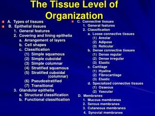
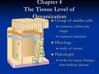
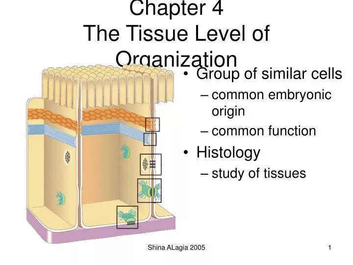
Chapter 4 The Tissue Level of Organization. Group of similar cells common embryonic origin common function Histology study of tissues. 4 Basic Tissues (1). Epithelial Tissue covers surfaces because cells are in contact lines hollow organs, cavities and ducts

E N D
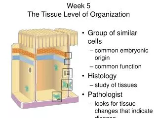
Chapter 4The Tissue Level of Organization • Group of similar cells • common embryonic origin • common function • Histology • study of tissues Shina ALagia 2005
4 Basic Tissues (1) • Epithelial Tissue • covers surfaces because cells are in contact • lines hollow organs, cavities and ducts • forms glands when cells sink under the surface • Connective Tissue • supports and binds structures together • stores energy as fat • provides immunity to disease Shina ALagia 2005
4 Basic Tissues (2) • Muscle Tissue • cells shorten in length producing movement • Nerve Tissue • cells that conduct electrical signals • detects changes inside and outside the body • responds with nerve impulses Shina ALagia 2005
Origin of Tissues • Primary germ layers within the embryo • endoderm • mesoderm • ectoderm • Tissue derivations • epithelium from all 3 germ layers • connective tissue & muscle from mesoderm • nerve tissue from ectoderm Shina ALagia 2005
Biopsy • Removal of living tissue for microscopic examination • Useful for diagnosis, especially cancer • Tissue preserved, sectioned and stained before microscopic viewing Shina ALagia 2005
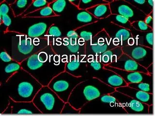
Epithelial Tissue -- General Features • Closely packed cells forming continuous sheets • Cells sit on basement membrane • Apical (upper) free surface • Avascular---without blood vessels • nutrients diffuse in from underlying connective tissue • Good nerve supply • Rapid cell division Shina ALagia 2005
Basement Membrane • Basal lamina • from epithelial cells • collagen fibers • Reticular lamina • secreted by connective tissue cells • reticular fibers • holds cells to connective tissue • guide for cell migration during development Shina ALagia 2005
Types of Epithelium • Covering and lining epithelium • epidermis of skin • lining of blood vessels and ducts • lining respiratory, reproductive, urinary & GI tract • Glandular epithelium • secreting portion of glands • thyroid, adrenal, and sweat glands Shina ALagia 2005
Classification of Epithelium • Classified by arrangement of cells into layers • simple = one cell layer thick • stratified = many cell layers thick • pseudostratified = single layer of cells where all cells don’t reach apical surface • nuclei at found at different levels so it looks multilayered • Classified by shape of surface cells • squamous =flat • cuboidal = cube-shaped • columnar = tall column • transitional = shape varies with tissue stretching Shina ALagia 2005
Simple Squamous Epithelium • Single layer of flat cells • lines blood vessels (endothelium), body cavities (mesothelium) • very thin --- controls diffusion, osmosis and filtration • nuclei centrally located • Cells in direct contact with each other Shina ALagia 2005
Section of intestinal showing serosa Surface view of lining of peritoneal cavity Examples of Simple Squamous Shina ALagia 2005
Simple Cuboidal Epithelium • Single layer of cube-shaped cells viewed from the side • Nuclei round and centrally located • Lines tubes of kidney • Absorption or secretion Shina ALagia 2005
Example of Simple Cuboidal • Sectional view of kidney tubules Shina ALagia 2005
Nonciliated Simple Columnar • Single layer rectangular cells • Unicellular glands =goblet cells secrete mucus • lubricate GI, respiratory, reproductive and urinary systems • Microvilli = fingerlike cytoplasmic projections • for absorption in GI tract (stomach to anus) Shina ALagia 2005
Ex. Nonciliated Simple Columnar • Section from small intestine Shina ALagia 2005
Ciliated Simple Columnar Epithelium • Single layer rectangular cells with cilia • Mucus from goblet cells moved along by cilia • found in respiratory system and uterine tubes Shina ALagia 2005
Ex. Ciliated Simple Columnar • Section of uterine tube Shina ALagia 2005
Stratified Squamous Epithelium • Several cell layers thick • Surface cells flat • Keratinized = surface cells dead and filled with keratin • skin (epidermis) • Nonkeratinized = no keratin in moist living cells at surface • mouth, vagina Shina ALagia 2005
Example of Stratified Squamous • Section of vagina Shina ALagia 2005
Stratified Cuboidal Epithelium • Multilayered • Surface cells cuboidal • rare (only found in sweat gland ducts & male urethra) Shina ALagia 2005
Stratified Columnar Epithelium • Multilayered • Surface cells columnar • Rare (very large ducts & part of male urethra) Shina ALagia 2005
Multilayered Surface cells varying in shape from round to flat if stretched Lines hollow organs that expand from within (urinary bladder) Transitional Epithelium Shina ALagia 2005
Single cell layer All cells attach to basement membrane but not all reach free surface Nuclei at varying depths Respiratory system, male urethra & epididymis Pseudostratified Columnar Shina ALagia 2005
Glandular Epithelium • Derived from epithelial cells that sank below the surface during development • Exocrine glands • cells that secrete---sweat, ear wax, saliva, digestive enzymes onto free surface of epithelial layer • connected to the surface by tubes (ducts) • unicellular glands or multicellular glands • Endocrine glands • secrete hormones into the bloodstream • hormones help maintain homeostasis Shina ALagia 2005
Connective Tissues • Cells rarely touch due to extracellular matrix • Matrix(fibers & ground substance secreted by cells • Consistency varies from liquid, gel to solid • Does not occur on free surface • Good nerve & blood supply except cartilage & tendons Shina ALagia 2005
Cell Types • Blast type cells = retain ability to divide & produce matrix (fibroblasts, chondroblasts, & osteoblasts) • Cyte type cells = mature cell that can not divide or produce matrix (chondrocytes & osteocytes) • Macrophages develop from monocytes • engulf bacteria & debris by phagocytosis • Plasma cells develop from B lymphocytes • produce antibodies that fight against foreign substances • Mast cells produce histamine that dilate small BV • Adipocytes (fat cells) store fat Shina ALagia 2005
Connective Tissue Ground Substance • Supports the cells and fibers • Helps determine the consistency of the matrix • fluid, gel or solid • Contains many large molecules • hyaluronic acid is thick, viscous and slippery • condroitin sulfate is jellylike substance providing support • adhesion proteins (fibronectin) binds collagen fibers to ground substance Shina ALagia 2005
Collagen (25% of protein in your body) tough, resistant to pull, yet pliable formed from the protein collagen Elastin (lungs, blood vessels, ear cartilage) smaller diameter fibers formed from protein elastin surrounded by glycoprotein (fibrillin) can stretch up to 150% of relaxed length and return to original shape Reticular (spleen and lymph nodes) thin, branched fibers that form framework of organs formed from protein collagen Types of Connective Tissue Fibers Shina ALagia 2005
Embryonic Connective Tissue:Mesenchyme • Irregularly shaped cells • In semifluid ground substance with reticular fibers • Gives rise to all other types of connective tissue Shina ALagia 2005
Mature Connective Tissue • Loose connective tissue • Dense connective tissue • Cartilage • Bone • Blood • Lymph Shina ALagia 2005
Loose Connective Tissues • Loosely woven fibers throughout tissues • Types of loose connective tissue • areolar connective tissue • adipose tissue • reticular tissue Shina ALagia 2005
Areolar Connective Tissue • Cell types = fibroblasts, plasma cells, macrophages, mast cells and a few white blood cells • All 3 types of fibers present • Gelatinous ground substance Shina ALagia 2005
Areolar Connective Tissue • Black = elastic fibers, • Pink = collagen fibers • Nuclei are mostly fibroblasts Shina ALagia 2005
Adipose Tissue • Peripheral nuclei due to large fat storage droplet • Deeper layer of skin, organ padding, yellow marrow • Reduces heat loss, energy storage, protection • Brown fat found in infants has more blood vessels and mitochondria and responsible for heat generation Shina ALagia 2005
Dense Connective Tissue • More fibers present but fewer cells • Types of dense connective tissue • dense regular connective tissue • dense irregular connective tissue • elastic connective tissue Shina ALagia 2005
Dense Regular Connective Tissue • Collagen fibers in parallel bundles with fibroblasts between bundles of collagen fibers • White, tough and pliable when unstained (forms tendons) • Also known as white fibrous connective tissue Shina ALagia 2005
Dense Irregular Connective Tissue • Collagen fibers are irregularly arranged (interwoven) • Tissue can resist tension from any direction • Very tough tissue -- white of eyeball, dermis of skin Shina ALagia 2005
Cartilage • Network of fibers in rubbery ground substance • Resilient and can endure more stress than loose or dense connective tissue • Types of cartilage • hyaline cartilage • fibrocartilage • elastic cartilage Shina ALagia 2005
Hyaline Cartilage • Bluish-shiny white rubbery substance • Chondrocytes sit in spaces called lacunae • No blood vessels or nerves so repair is very slow • Reduces friction at joints as articular cartilage Shina ALagia 2005
Fibrocartilage • Many more collagen fibers causes rigidity & stiffness • Strongest type of cartilage (intervertebral discs) Shina ALagia 2005
Elastic Cartilage • Elastic fibers help maintain shape after deformations • Ear, nose, vocal cartilages Shina ALagia 2005
Growth & Repair of Cartilage • Grows and repairs slowly because is avascular • Interstitial growth • chondrocytes divide and form new matrix • occurs in childhood and adolescence • Appositional growth • chondroblasts secrete matrix onto surface • produces increase in width Shina ALagia 2005
Spongy bone sponge-like with spaces and trabeculae trabeculae = struts of bone surrounded by red bone marrow no osteons (cellular organization) Compact bone solid, dense bone basic unit of structure is osteon (haversian system) Protects, provides for movement, stores minerals, site of blood cell formation Bone (Osseous) Tissue Shina ALagia 2005
Compact Bone • Osteon = lamellae (rings) of mineralized matrix • calcium & phosphate---give it its hardness • interwoven collagen fibers provide strength • Osteocytes in spaces (lacunae) in between lamellae • Canaliculi (tiny canals) connect cell to cell Shina ALagia 2005
Blood • Connective tissue with a liquid matrix = the plasma • Cell types = red blood cells (erythrocytes), white blood cells (leukocytes) and cell fragments called platelets • Provide clotting, immune functions, carry O2 and CO2 Shina ALagia 2005
Lymph • Interstitial fluid being transported in lymphatic vessels • Contains less protein than plasma • Move cells and substances (lipids) from one part of the body to another Shina ALagia 2005
Membranes • Epithelial layer sitting on a thin layer of connective tissue (lamina propria) • Types of membranes • mucous membrane • serous membrane • synovial membrane • cutaneous membrane (skin) Shina ALagia 2005
Mucous Membranes • Lines a body cavity that opens to the outside • mouth, vagina, anus etc • Epithelial cells form a barrier to microbes • Tight junctions between cells • Mucous is secreted from underlying glands to keep surface moist Shina ALagia 2005
Serous Membranes • Simple squamous cells overlying loose CT layer • Squamous cells secrete slippery fluid • Lines a body cavity that does not open to the outside such as chest or abdominal cavity • Examples • pleura, peritoneum and pericardium • membrane on walls of cavity = parietal layer • membrane over organs in cavity = visceral layer Shina ALagia 2005
Synovial Membranes • Line joint cavities of all freely movable joints • No epithelial cells---just special cells that secrete slippery fluid Shina ALagia 2005