Download
1 / 69
710 likes | 926 Views
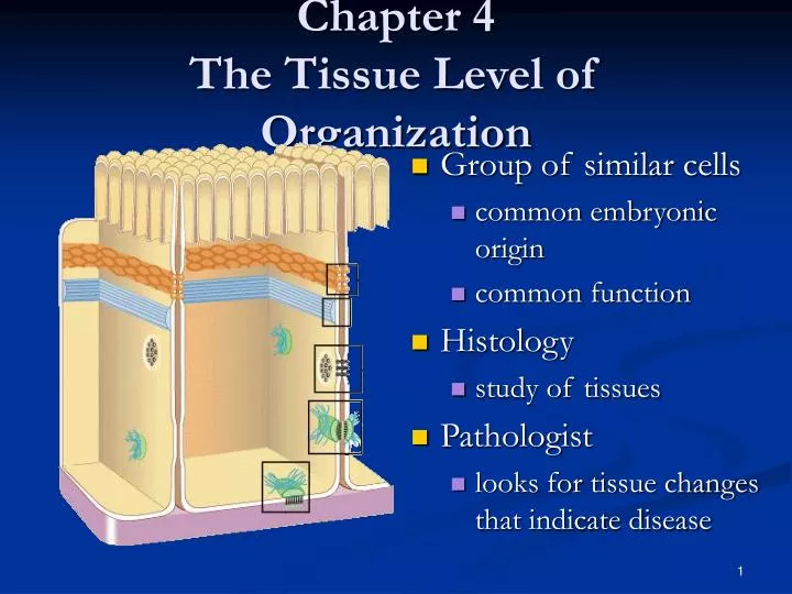
Chapter 4 The Tissue Level of Organization. Group of similar cells common embryonic origin common function Histology study of tissues Pathologist looks for tissue changes that indicate disease. 4 Basic Tissues (1). Epithelial Tissue covers surfaces because cells are in contact

E N D
Chapter 4The Tissue Level of Organization • Group of similar cells • common embryonic origin • common function • Histology • study of tissues • Pathologist • looks for tissue changes that indicate disease
4 Basic Tissues (1) • Epithelial Tissue • covers surfaces because cells are in contact • lines hollow organs, cavities and ducts • forms glands when cells sink under the surface • Connective Tissue • material found between cells • supports and binds structures together • stores energy as fat • provides immunity to disease
4 Basic Tissues (2) • Muscle Tissue • cells shorten in length producing movement • Nerve Tissue • cells that conduct electrical signals • detects changes inside and outside the body • responds with nerve impulses
Origin of Tissues • Primary germ layers within the embryo • endoderm • mesoderm • ectoderm • Tissue derivations • epithelium from all 3 germ layers • connective tissue & muscle from mesoderm • nerve tissue from ectoderm
Biopsy • Removal of living tissue for microscopic examination • surgery • needle biopsy • Useful for diagnosis, especially cancer • Tissue preserved, sectioned and stained before microscopic viewing
Cell Junctions • Tight junctions • Adherens junctions • Gap junctions • Desmosomes • Hemidesmosomes
Tight Junctions • Watertight seal between cells • Plasma membranes fused with a strip of proteins • Common between cells that line GI and bladder
Adherens Junctions • Holds epithelial cells together • Structural components • plaque = dense layer of proteins inside the cell membrane • microfilaments extend into cytoplasm
Desmosomes • Resists cellular separation and cell disruption • Cellular support of cardiac muscle
Hemidesmosomes • Half a desmosome • Connect cells to extracellular material • basement membrane
Gap Junctions • Tiny space between plasma membranes of 2 cells • Crossed by protein channels called connexons forming fluid filled tunnels • Cell communication with ions & small molecules • Muscle and nerve impulses spread from cell to cell • heart and smooth muscle of gut
Epithelial Tissue -- General Features • Closely packed cells forming continuous sheets • Cells sit on basement membrane • Apical (upper) free surface • Avascular---without blood vessels • nutrients diffuse in from underlying connective tissue • Good nerve supply • Rapid cell division • Covering / lining versus glandular types
Basement Membrane • Basal lamina • from epithelial cells • collagen fibers • Reticular lamina • secreted by connective tissue cells • reticular fibers • holds cells to connective tissue • guide for cell migration during development
Types of Epithelium • Covering and lining epithelium • epidermis of skin • lining of blood vessels and ducts • lining respiratory, reproductive, urinary & GI tract • Glandular epithelium • secreting portion of glands • thyroid, adrenal, and sweat glands
Classification of Epithelium • Classified by arrangement of cells into layers • simple = one cell layer thick • stratified = many cell layers thick • pseudostratified = single layer of cells where all cells don’t reach apical surface • nuclei at found at different levels so it looks multilayered • Classified by shape of surface cells • squamous =flat • cuboidal = cube-shaped • columnar = tall column • transitional = shape varies with tissue stretching
Simple Squamous Epithelium • Single layer of flat cells • lines blood vessels (endothelium), body cavities (mesothelium) • very thin --- controls diffusion, osmosis and filtration • nuclei centrally located • Cells in direct contact with each other
Section of intestinal showing serosa Surface view of lining of peritoneal cavity Examples of Simple Squamous
Simple Cuboidal Epithelium • Single layer of cube-shaped cells viewed from the side • Nuclei round and centrally located • Lines tubes of kidney • Absorption or secretion
Example of Simple Cuboidal • Sectional view of kidney tubules
Nonciliated Simple Columnar • Single layer rectangular cells • Unicellular glands =goblet cells secrete mucus • lubricate GI, respiratory, reproductive and urinary systems • Microvilli = fingerlike cytoplasmic projections • for absorption in GI tract (stomach to anus)
Ex. Nonciliated Simple Columnar • Section from small intestine
Ciliated Simple Columnar Epithelium • Single layer rectangular cells with cilia • Mucus from goblet cells moved along by cilia • found in respiratory system and uterine tubes
Ex. Ciliated Simple Columnar • Section of uterine tube
Stratified Squamous Epithelium • Several cell layers thick • Surface cells flat • Keratinized = surface cells dead and filled with keratin • skin (epidermis) • Nonkeratinized = no keratin in moist living cells at surface • mouth, vagina
Example of Stratified Squamous • Section of vagina
Papanicolaou Smear (Pap smear) • Collect sloughed off cells of uterus and vaginal walls • Detect cellular changes (precancerous cells) • Annually for women over 18 or if sexually active
Stratified Cuboidal Epithelium • Multilayered • Surface cells cuboidal • rare (only found in sweat gland ducts & male urethra)
Stratified Columnar Epithelium • Multilayered • Surface cells columnar • Rare (very large ducts & part of male urethra)
Multilayered Surface cells varying in shape from round to flat if stretched Lines hollow organs that expand from within (urinary bladder) Transitional Epithelium
Single cell layer All cells attach to basement membrane but not all reach free surface Nuclei at varying depths Respiratory system, male urethra & epididymis Pseudostratified Columnar
Glandular Epithelium • Derived from epithelial cells that sank below the surface during development • Exocrine glands • cells that secrete---sweat, ear wax, saliva, digestive enzymes onto free surface of epithelial layer • connected to the surface by tubes (ducts) • unicellular glands or multicellular glands • Endocrine glands • secrete hormones into the bloodstream • hormones help maintain homeostasis
Structural Classification of Exocrine Glands • Unicellular are single-celled glands • goblet cells • Multicellular glands • branched (compound) or unbranched (simple) • tubular or acinar (flask-like) shape
Methods of Glandular Secretion • Merocrine -- most glands • cells release their products by exocytosis---saliva, digestive enzymes & sweat • Apocrine • smelly sweat & milk • upper part of cell possibly pinches off & dies (perhaps--see EM data) • Holocrine -- oil gland • whole cells die & rupture to release their products
Connective Tissues • Cells rarely touch due to extracellular matrix • Matrix(fibers & ground substance secreted by cells • Consistency varies from liquid, gel to solid • Does not occur on free surface • Good nerve & blood supply except cartilage & tendons
Cell Types • Blast type cells = retain ability to divide & produce matrix (fibroblasts, chondroblasts, & osteoblasts) • Cyte type cells = mature cell that can not divide or produce matrix (chondrocytes & osteocytes) • Macrophages develop from monocytes • engulf bacteria & debris by phagocytosis • Plasma cells develop from B lymphocytes • produce antibodies that fight against foreign substances • Mast cells produce histamine that dilate small BV • Adipocytes (fat cells) store fat
Connective Tissue Ground Substance • Supports the cells and fibers • Helps determine the consistency of the matrix • fluid, gel or solid • Contains many large molecules • hyaluronic acid is thick, viscous and slippery • condroitin sulfate is jellylike substance providing support • adhesion proteins (fibronectin) binds collagen fibers to ground substance
Collagen (25% of protein in your body) tough, resistant to pull, yet pliable formed from the protein collagen Elastin (lungs, blood vessels, ear cartilage) smaller diameter fibers formed from protein elastin surrounded by glycoprotein (fibrillin) can stretch up to 150% of relaxed length and return to original shape Reticular (spleen and lymph nodes) thin, branched fibers that form framework of organs formed from protein collagen Types of Connective Tissue Fibers
Marfan Syndrome • Inherited disorder of fibrillin gene • Abnormal development of elastic fibers • Tendency to be tall with very long legs, arms, fingers and toes • Life-threatening weakening of aorta may lead to rupture
Mature Connective Tissue • Loose connective tissue • Dense connective tissue • Cartilage • Bone • Blood • Lymph
Loose Connective Tissues • Loosely woven fibers throughout tissues • Types of loose connective tissue • areolar connective tissue • adipose tissue • reticular tissue
Areolar Connective Tissue • Cell types = fibroblasts, plasma cells, macrophages, mast cells and a few white blood cells • All 3 types of fibers present • Gelatinous ground substance
Areolar Connective Tissue • Black = elastic fibers, • Pink = collagen fibers • Nuclei are mostly fibroblasts
Adipose Tissue • Peripheral nuclei due to large fat storage droplet • Deeper layer of skin, organ padding, yellow marrow • Reduces heat loss, energy storage, protection • Brown fat found in infants has more blood vessels and mitochondria and responsible for heat generation
Liposuction or Suction Lipectomy • Suctioning removal of subcutaneous fat for body contouring • Dangers include fat emboli, infection, injury to internal organs and excessive pain
Reticular Connective Tissue • Network of fibers & cells that produce framework of organ • Holds organ together (liver, spleen, lymph nodes, bone marrow)
Cartilage • Network of fibers in rubbery ground substance • Resilient and can endure more stress than loose or dense connective tissue • Types of cartilage • hyaline cartilage • fibrocartilage • elastic cartilage
Hyaline Cartilage • Bluish-shiny white rubbery substance • Chondrocytes sit in spaces called lacunae • No blood vessels or nerves so repair is very slow • Reduces friction at joints as articular cartilage
Fibrocartilage • Many more collagen fibers causes rigidity & stiffness • Strongest type of cartilage (intervertebral discs)
Elastic Cartilage • Elastic fibers help maintain shape after deformations • Ear, nose, vocal cartilages
Growth & Repair of Cartilage • Grows and repairs slowly because is avascular • Interstitial growth • chondrocytes divide and form new matrix • occurs in childhood and adolescence • Appositional growth • chondroblasts secrete matrix onto surface • produces increase in width