Download
1 / 130
1.37k likes | 1.93k Views
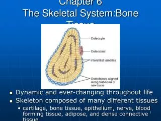
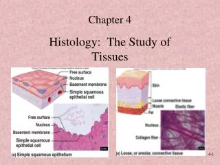
Chapter 4: The Tissue Level of Organization. Introduction. Tissue: Collection of specialized cells that perform limited number of functions Histology: The study of tissues What are the four tissues of the body?. The four tissues of the Body. Epithelial Tissue: “covering”

E N D
Introduction • Tissue: • Collection of specialized cells that perform limited number of functions • Histology: • The study of tissues What are the four tissues of the body?
The four tissues of the Body • Epithelial Tissue: “covering” • Covers exposed surfaces • Lines internal passageways • Forms glands • Connective Tissue: “support” • Fills internal spaces • Provides structure and strength to support other tissues • Transports materials • Stores energy
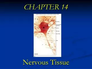
The four tissues of the Body • Muscle Tissue: “movement” • Specialized for contraction • Skeletal muscle, heart muscle, and walls of hollow organs • Neural Tissue: “control” • Carries electrical signals from 1 part of the body to another
Primary Germ Layer • Embryonic layers give rise to all four tissue types in adults • Ectoderm: nervous, epithelial (epidermis) • Mesoderm: muscle, connective, epithelial (endothelium + mesothelium) • Endoderm: epithelial (mucosa)
KEY CONCEPT • Tissues are collections of cells and cell products that perform specific, limited functions • 4 tissue types form all the structures of the human body: • epithelial, connective, muscle, and neural
Epithelial Tissues • 2 categories: • Epithelia: • layers of cells covering internal or external surfaces • Glands: • structures that produce fluid secretions
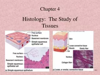
Characteristics of Epithelia Structures of Epithelia: • Cellularity: • little extracellular matrix, mostly cells • Contacts: • cells linked by tight junctions • Polarity: • apical + basal surfaces, separate functions • Attachment: • attached to connective tissue (CT) via basal lamina • Avascularity: • diffusion of CT • Regeneration: • high turnover, stem cells at basal surface
Characteristics of Epithelia Functions of Epithelia: • Provide physical protection: • abrasion, dehydration, infection • Control permeability: • semi-permeability, covers all surfaces • Provide sensation: • sensory neurons • Produce specialized secretions (glandular epithelium): • protection, chemical messengers
Free Surface and Attached Surface 1. Apical Surface:exposed to environment, may have: • Microvilli: absorption or secretion • Cilla: fluid movement 2. Basolateral Surface: attachmenttoneighboring cells via intercellular connections Figure 4–1
Intercellular Connections • Support and communication • General Adhesion: Large Connections 1. CAMs (cell adhesion molecules): • Connect adjacent membranes or binds extracellular materials (e.g. basal lamina) 2. Intercellular cement: • Thin layer of hyaluronan (proteoglycan): • Attach adjacent membranes • Specific Adhesion=Cell Junctions • Tight Junctions • Gap Junctions • Desmosomes
Cell Junctions • Form bonds with other cells or extracellular material: • Tight Junctions: • interlocking proteins, bind lipid portion of membrane, water tight seal • Gap Junctions: • connexons form channel, allow molecules to pass for communication • Desmosomes: • CAMs + intercellular cement on dense area attached to cytoskeleton, resist stretching and twisting
Between 2 cell membranes interlocking proteins, bind lipid portion of membrane, water tight seal Prevents passage of water and solutes Tight Junctions Figure 4–2b
Connexons form protein channels, allow molecules to pass for communication Rapid communication Allow ions to pass Coordinated contractions in heart muscle Gap Junctions Figure 4–2c
Cell Adhesion Molecules + intercellular cement on dense area are attached to the cytoskeleton - resist stretching and twisting Belt Desmosomes: -continuous band in apical region, attached to microfilaments Button Desmosomes: - “spot weld”, attached to intermediate filaments 3. Hemidesmosomes: - Half button desmosome at basal surface, attaches to basal lamina Desmosomes Figure 4–2d
Basal Lamina (a.k.a. basement membrane) • Lamina lucida: • Produced by epithelia • Glycoproteins + fine filaments restrict large molecule movement • Lamina densa: • Produced by connective tissue • Coarse protein fibers • Provides strength and filtration
Repairing and Replacing Epithelia • Epithelia stem cells are anchored to lamina lucida • Epithelia are replaced by division of germinative cells (stem cells) • Stem cells divide and migrate toward apical region
Classes of Epithelia • Based on shape and layers • Shape: (all are hexagonal from the top) • Squamous: flat, disc shaped nucleus • Cuboidal: cube or square, center round nucleus • Columnar: tall, basal oval nucleus Table 4–1
Layers • Simple epithelium: • single layer of cells • Function: • absorption, secretion, filtration • Stratified epithelium: • 2 or more layers of cells • Function: • Protection **In stratified, name for apical cell shape**
Simple squamous epithelium Stratified squamous epithelium Simple cuboidal epithelium Stratified cuboidal epithelium Transitional epithelium Simple columnar epithelium Pseudostratified columnar epithelium Stratified columnar epithelium Eight Types of Epithelial Tissues
1. Simple Squamous Epithelium • Thin, delicate • Locations: found in protected regions • Mesothelium (serosa), endothelium (blood vessels, heart), kidney tubules, cornea, and alveoli of lungs • Functions: • Absorption, diffusion, filtration, or secretion Figure 4–3a
2. Stratified Squamous Epithelium • Basal cells look cuboidal, apical cells squamous • Found on exposed surfaces • Functions: provide protection from abrasion, pathogens, and chemicals • Two types: • Nonkeratinized = mucosa - Kept moist - All cells are nucleated Location: mouth, esophagus, anus, and vagina • Keratinized = epidermis - dry, apical cells dead - cells contain keratin protein to resist dehydration and adds strength Figure 4–3b
3. Simple Cuboidal Epithelia • Location: * Functions: • Kidney tubules - Secretion • Pancreas -Absorption • Salivary glands • Thyroid
4. Stratified Cuboidal Epithelium • Rare • Typically 2 layers • Location: * Function: • Some sweat glands - Secretion • Some mammary glands - Absorption Figure 4–4b
5. Transitional Epithelium • Relaxed: looks like stratified cuboidal • Stretched: looks squamous • Location: * Function • Urinary bladder - tolerate excessive stretching • Ureters Figure 4–4c
Columnar Epithelia • Simple columnar epithelium: • absorption and secretion • Pseudostratified columnar epithelium: • cilia movement • Stratified columnar epithelium: • protection
6. Simple Columnar Epithelium • Nuclei line up near the basal lamina • Apical surface of cells often has microvilli = “brush border” (in intestine) • Goblet cells often present: secrete mucus • Locations: • Stomach, intestine, gall bladder, uterine tubes, and collecting ducts of kidney • Functions: • Absorption or secretion Figure 4–5a
7. Pseudostratified Columnar Epithelium • Several cell types: varying shapes and functions • All cells contact basal lamina • Some too short to reach apical surface • Nuclei scattered so it appears stratified • Tall cells have cilia on apical surface • Goblet cells (mucus) often present • Location: Function: • Nasal cavity, trachea, bronchi - move material • Male reproductive tract across surface • Female uterine tubes Figure 4–5b
8. Stratified Columnar Epithelium • Rare • Two layers or multiple layers with only apical layer columnar • Locations (tiny parts of): • Pharynx, epiglottis, anus, mammary glands, salivary glands, and urethra • Functions: • Minor protection Figure 4–5c
Glandular Epithelia For secretion, makes up glands • Endocrine glands: “internally secreting” -secrete into interstital fluid blood -secretions = hormones -regulate and coordinate activities e.g. pancreas, thyroid, thymus, pituitary • Exocrine glands: “externally secreting” -secrete into duct epithelial surface -e.g. digestive enzymes, perspiration, tears, milk, and mucus -Classified three ways: 1. Mode of Secretion 2. Type of Secretion 3. Structure
A. Modes of Secretion 1. Merocrine secretion: -product released from secretory vesicles by exocytosis -e.g. mucus, sweat • Apocrine Secretion: - product accumulates in vesicles - apical region of cell which vesicles is shed to release product -e.g. milk Figure 4–6a
Modes of Secretion 3. Holocrine secretion - product accumulates in vesicles - whole cell is lysed to release product - cell dies, must be replaced by stem cells e.g. sebum Figure 4–6c
B. Types of Secretion • Serous Glands: water + enzymes - e.g. parotid salivary gland • Mucus Glands: mucin (+water = mucus) - e.g. goblet cell • Mixed exocrine glands: (serous + mucus secretion) -e.g. submandibular salivary gland
C. Gland Structure • Exocrine glands can be classified as: • unicellular glands: 1 cell • E.g. goblet cell which are scattered among epithelia • Found in intestinal lining • multicellular glands: group of cells named for shape and structure
Structure of Multicellular Exocrine Glands • Structural classes of exocrine glands Simple Glands = undivided tube shape blind pockets chamberlike Figure 4–7 (1 of 2)
Structure of Multicellular Exocrine Glands Compound Glands = Divided tube shaped blind pockets chamberlike Figure 4–7 (2 of 2)
Which of the following is NOT a characteristic of epithelial tissue? A.It is composed entirely of cells. B. It stores energy reserves. C. It is avascular. D. It is capable of regeneration.
An epithelial surface bears many microvilli. What is the probable function of this epithelium? A.absorption B. secretion C. transportation D. sensation
What is the functional significance of gap junctions? A.They maintain water-tight passages. B. They resist stretching and twisting. C. They coordinate the function of tissue. D. They attach cells to extracellular matrix.
Using a light microscope, you examine a tissue and see a simple squamous epithelium on the outer surface. Can this be a sample of the skin surface? A.Yes B.No
Why do the pharynx, esophagus, anus, and vagina have the same epithelial organization? A.All are subject to mechanical trauma. B. All are subject to abrasion. C. All must be able to expand. D. A and B are correct.
The secretory cells of sebaceous glands fill with secretions and then rupture, releasing their contents. Which type of secretion is this? A.acinar B. apocrine C. merocrine D. holocrine
A gland has no ducts to carry the glandular secretions, and the gland’s secretions are released directly into the extracellular fluid. Which type of gland is this? A.exocrine gland B. endocrine gland C. acinar gland D. tubular gland
Structures and functions of different types of Connective Tissues.
Connective Tissues • Features: • Never exposed to the environment • Usually vascularized • Consists of cells in a matrix • Components: • Specialized cells: • Produce matrix, provide protection 2. Extracellular protein fibers: - Support, strength 3. Ground Substance: - gel fluid, consists of: * interstitial fluid, cell adhesion molecules, and GAGs (glycosaminoglycans) - proteoglycans that form a gel Fibers + Ground Substance = Matrix
The Matrix • The extracellular components of connective tissues (fibers and ground substance): • majority of cell volume • determines specialized function
Functions of Connective Tissue • Establish structural framework • Transport fluid and dissolved materials • Protect organs • Support, surround, interconnect tissues • Store energy reserves • Insulate body • Defend against pathogens
Classification of Connective Tissues • Connective tissue proper: • Many cell types and fiber types in thick ground substance • Loose: open fiber framework • Dense: tightly packed fibers • Fluid connective tissues: • May cell types in watery matrix with soluble fibers • Supportive connective tissues: • Limited cell population in tightly packed matrix
Connective Tissues Derived from Mesenchyme Embryonic CT: - mesenchymal cells in gelatinous matrix with fine fibers