Download
1 / 97
1.07k likes | 1.61k Views
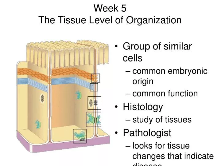
Week 5 The Tissue Level of Organization. Group of similar cells common embryonic origin common function Histology study of tissues Pathologist looks for tissue changes that indicate disease. 4 Basic Tissues (1). Epithelial Tissue covers exposed surfaces ( skin )

E N D
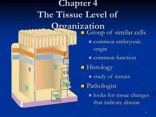
Week 5 The Tissue Level of Organization • Group of similar cells • common embryonic origin • common function • Histology • study of tissues • Pathologist • looks for tissue changes that indicate disease
4 Basic Tissues (1) • Epithelial Tissue • covers exposed surfaces ( skin ) • lines hollow organs, internal passageways and ducts ( digestive tract, urinary tract, reproductive) • forms glands- when cells sink under the surface • Connective Tissue • material found between cells • supports and binds structures together • stores energy as fat • provides immunity to disease
4 Basic Tissues (2) • Muscle Tissue • cells shorten in length producing movement • Nerve Tissue • cells that conduct electrical signals • detects changes inside and outside the body • responds with nerve impulses
Biopsy • Removal of living tissue for microscopic examination • surgery • needle biopsy • Useful for diagnosis, especially cancer • Tissue preserved, sectioned and stained before microscopic viewing
Maintaining the integrity of Epithelium To be effective as a barrier, an epithelium must form a complete cover or lining. 3 factors help maintain the integrity of the epithelium • Intercellular connection -Cell adhesion molecule, cell junctions • Attachment to basement membrane – 2 layers, clear and dense layer • Maintenance and repair –epithelium live hard lives. Exposed to enzymes, toxic chemicals, bacteria, and mechanical abrasions. Stem cells called GERMATIVE cells divide continually.
Cell Junctions • Tight junctions • Adherens junctions • Gap junctions • Desmosomes • Hemidesmosomes
Tight Junctions • Watertight seal between cells. Preventing passage of water and solutes • Plasma membranes fused with a strip of proteins • Common between cells that line GI and bladder • Don’t want acids, enzymes and waste leaking.
Adherens Junctions • Holds epithelial cells together • Structural components • plaque = dense layer of proteins inside the cell membrane • microfilaments extend into cytoplasm • integral membrane proteins connect to membrane of other cell
Desmosomes • Resists cellular separation, cell disruption, twisting, compression. • Similar structure to adherens junction except intracellular intermediate filaments cross cytoplasm of cell • Found in superficial layer of skin. (Skin peels when damages.)
Gap Junctions • Tiny space between plasma membranes of 2 cells • Crossed by protein channels called connexons forming fluid filled tunnelsallowing sugars, amino acids, and electrolytes to pass. • Cell communication rapidly with ions & small molecules • Muscle and nerve impulses spread from cell to cell • heart and smooth muscle of gut
Epithelial Tissue -- General Features • Closely packed cells forming continuous sheets held together by cell junctions. • Cells sit on basement membrane • Apical (upper) free surface • Avascular---without blood vessels • nutrients diffuse in from underlying connective tissue • Good nerve supply • Rapid cell division if damagedvia stem cells • Covering- line all passage ways that come into contact with outside world. Skin, Digestive, respiratory, reproductive, urinary, brain, blood vessels, heart.
Functions • Physical protection – protects exposed and internal surfaces from abrasions, dehydration, and destruction from chemical agents. • Absorption – The lining of the gut and respiratory tract allow for nutrients to be absorbed from the gut and the exchange of gases between air in lungs and blood. • Excretion- The unique lining of the kidney tubules makes the excretion and concentration of excretory products in the urine possible. • Produces secretions – Glandular epithelial is adapted for secretory activity (hormones, mucus, digestive juices, and sweat) • Provides sensation – good nerve supply- sensory nerves of eyes, ears, nose, skin. • Specialized Epithelium – a) Microvilli – absorption/secretion – found in digestive and urinary tracts b) Cilia – Respiratory tracts, the synchronized beating moves mucous up from lungs to throat.
Basement Membrane • Basal lamina • from epithelial cells • collagen fibers • Reticular lamina • secreted by connective tissue cells • reticular fibers-strength • holds cells to connective tissue • guide for cell migration during development
Types of Epithelium • Covering and lining epithelium • epidermis of skin • lining of blood vessels and ducts • lining respiratory, reproductive, urinary & GI tract • Glandular epithelium • secreting portion of glands • thyroid, adrenal, and sweat glands
Classification of Epithelium • Classified by arrangement of cells into layers • simple=one cell layer thick. Secretion or absorption (intestine ,lungs) • stratified = many cell layers thick. Located where protection from mechanical or chemical stresses are needed. ( skin , mouth ) • pseudostratified = single layer of cells where all cells don’t reach apical surface • nuclei at found at different levels so it looks multilayered • Classified by shape of surface cells • squamous =flat • cuboidal = cube-shaped • columnar = tall column • transitional = shape varies with tissue stretching
Simple Squamous Epithelium • Single layer of flat cells • lines blood vessels(endothelium heart chambers),body cavities(mesothelium :pleura, pericardium, peritoneum) • very thin --- controls diffusion, osmosis and filtration • nuclei centrally located • Cells in direct contact with each other • Good for gas exchange at lungs(alveoli), absorption at intestines, and re-absorption loop of henley at kidneys
Examples of Simple Squamous • Section of intestinal showing serosa • Surface view of lining of peritoneal cavity
Simple Cuboidal Epithelium • Single layer of cube-shaped cells viewed from the side • Nuclei round and centrally located • Lines tubes of kidney – proximal and distal tubules • Absorption or secretion
Example of Simple Cuboidal • Sectional view of kidney tubules
Nonciliated Simple Columnar • Single layer rectangular cells • Unicellular glands =goblet cells secrete mucus • lubricate GI, respiratory, reproductive and urinary systems • Microvilli = fingerlike cytoplasmic projections • for absorption in GI tract (stomach to anus)
Ex. Nonciliated Simple Columnar • Section from small intestine
Ciliated Simple Columnar Epithelium • Single layer rectangular cells with cilia • Mucus from goblet cells moved along by cilia • found in respiratory system and uterine tubes
Ex. Ciliated Simple Columnar • Section of uterine tube
Stratified Squamous Epithelium • Several cell layers thick • Surface cells flat. Located where mechanical and chemical stresses are severe • Keratinized = surface cells dead and filled with keratin. This layer is tough and water resistant. • skin (epidermis) • Nonkeratinized- resists abrasions, but will dry out if not kept moist. No keratin in moist living cells at surface • mouth, vagina
Example of Stratified Squamous • Section of vagina
Papanicolaou Smear (Pap smear) • Collect sloughed off cells of uterus and vaginal walls • Detect cellular changes (precancerous cells) • Annually for women over 18 or if sexually active • HPV – 80 % of women over 50 have it. • Low risk- irregular cells, high risk- leads to cervix cancer. • Guardasil – protects only 4 out of 10 strains.
Stratified Cuboidal Epithelium • Multilayered • Surface cells cuboidal • rare (only found in sweat gland ducts & male urethra)
Stratified Columnar Epithelium • Multilayered • Surface cells columnar • Rare (very large ducts & part of male urethra)
Transitional Epithelium • Multilayered • Surface cells varying in shape from round to flat if stretched • Lines hollow organs that expand from within (urinary bladder)
Pseudostratified Columnar • Single cell layer • All cells attach to basement membrane but not all reach free surface • Nuclei at varying depths • Respiratory system, male urethra & epididymis
Glandular Epithelium • Derived from epithelial cells that sank below the surface during development • Exocrine glands • cells that secrete---sweat, tears, milk, ear wax, saliva, digestive enzymes onto free surface of epithelial layer • connected to the surface by tubes (ducts) • Endocrine glands – ductless glands (thyroid, pituitary, adrenal glands) • secrete hormones into the bloodstream • hormones help maintain homeostasis • Sjorgens syndrome – autoimmune disease of exocrine glands. Dry eyes, mouth
Methods of Glandular Secretion • Merocrine (part)-- Most glands • cells release their products by exocytosis---saliva, digestive enzymes & sweat • Apocrine (off) • milk • upper part of cell possibly pinches off & dies • Holocrine (entire) -- oil gland • whole cells die & rupture to release their products
Exocrine glands- 3 types • 1) Serous Glands – Secretes a watery solution that contains enzymes. Parotid gland. • 2) Mucous glands –secrete mucins that hydrate to form mucous. Salivary glands, submucosal glands of the small intestine. • 3) Mixed glands- submandibular
Week 5 Connective Tissues • Cells rarely touch due to extracellular matrix • Matrix (fibers & ground substance secreted by cells • Consistency varies from liquid, gel to solid • Good nerve & blood supply except cartilage & ligaments)
Extracellular Matrix The ties that bind us together –healthy ECM means reduced pain/degenerative changes and increased function
Down-regulate the expression collagen-damaging enzymes (MMPs) to support balanced healing of extracellular matrix tissues Support for Extra-Cellular (Connective) Tissues Unhealthy ECM is found in patients that: • Are suffering from soft-tissue injury • Routinely suffer from repetitive injury • Have difficulty maintaining alignment • Are experiencing degradation of connective tissues
Connective Tissues = Extracellular Matrix tendons joints ligaments cartilage fascia http://images.google.com/imgres?imgurl=http://www.pride.hofstra.edu/~akroto1/img009.jpg&imgrefurl=http://www.pride.hofstra.edu/~akroto1/bioengg.html&h=450&w=600&sz=75&hl=en&start=3&tbnid=9BMB7hzszEIkUM:&tbnh=101&tbnw=135&prev=/images%3Fq%3Dextracellular%26gbv%3D2%26hl%3Den
Extracellular matrix is the structural scaffold for the cells and is the defining feature of connective tissue Non-cellular components • Heparan sulfate • Chondroitin sulfate • Keratan sulfate • Laminin • Hyaluronic Acid • Collagen • Fibronectin • Elastin • Cellular Components • Fibroblasts • Chondrocytes • Osteoblasts
A complex web that of connective tissues that • Provide adhesion to cells • Act as a structural scaffold • Actively participate in intracellular signaling • Translates mechanical loading into cellular response The extracellular matrix is composed of tissues that serve multiple purposes “The ECM is a conglomerate of substances in which biochemicals and biophysical properties allow for the construction of a flexible network that integrates information from loading and converts it into mechanical capacities…It serves as a scaffold for adhesion of cells mediated by integrins, dystroglycan, and proteoglycans at the cell surface and of tyrosine kinase receptors. The interaction between the ECM and the adhesion molecules leads to activation of intracellular signaling pathways and cytoskeletal rearrangement.” Kjaer, M. Physiol. Rev. 84:649-698, 2004. http://content.answers.com/main/content/img/McGrawHill/Encyclopedia/images/CE157300FG0010.gif
Extracellular matrix tissues are degraded after injury physiology of an injury • Prostaglandins and thromboxanes are created which signal pain and induce inflammation • Chondrocytes in the extracellular matrix release matrix metalloproteinase enzymes (MMPs) into the area of injury • MMPs degrade collagens, elastins, basement membranes
“MMP-1 and MMP-13 are collagenolytic MMPs that have been most strongly associated with cartilage collagenolysis” • “chondrocytes are stimulated to secrete elevated levels of MMPs that, once activated, mediate the proteolysis of tendon, bone and cartilage.” Down-regulate the expression of MMP-13 to support connective tissue Litherland et al., JBC, Papers in press, March 10, 2008
MMP-13 MMP-13 MMP-13 MMPs further degrade collagen tissue during injury or inflammation MMP-9 MMP- 2 MMP-1 Modulate expression of MMPs to support healthy remodeling of connective tissue MMPs are released from the chondrocyte in response to injury or inflammation Matrix metalloproteinases digest collagen and connective tissue Chondrocyte Collagen Triple Helix
Clinical conditions associated with increased expression of MMPs Periodontal disease Tendon injury Fracture healing MMPs Degenerative discs Wound repair
“Matrix metalloproteinase inhibitors may play a supportive role in the treatment of tendinopathy by limiting the MMP-mediated degradation of the extracellular matrix.” MMPs are a primary target to address tendonitis and connective tissue disorder Am J Sports Med. 2007 May;35(5):763-9. Epub 2007 Feb 9. Links Matrix metalloproteinase inhibitors prevent a decrease in the mechanical properties of stress-deprived tendons: an in vitro experimental study. Arnoczky SP,Lavagnino M,Egerbacher M, Caballero O, Gardner K. “An increase in matrix metalloproteinases (MMPs) and the resulting degradation of the extracellular matrix have been implicated in the pathogenesis of tendinopathy. Matrix metalloproteinase inhibitors prevented the activation of MMP-13 and significantly inhibited pericellular matrix degeneration and the loss of material properties associated with stress deprivation. Matrix metalloproteinase inhibitors may play a supportive role in the treatment of tendinopathy by limiting the MMP-mediated degradation of the extracellular matrix.”
Nutritional strategy to balance the expression of MMPs • Hops & Berberine to modulate MMP-13 • Selenium to address MMP-1 & MMP-2 • Folic acid to impact MMP-9 • Niacinamide to address tissue-damaging PARS Poly(ADP-ribose) polymerase inhibition reduces TNF-induced inflammatory response in rheumatoid synovial fibroblasts. García S, Bodaño A, Pablos JL, Gómez-Reino JJ, Conde C. “We show, for the first time, that PARP inhibition either with specific inhibitors or by siRNA transfection significantly reduced TNF-induced cytokine and chemokine expression in fibroblast like synoviocytes cells from RA patients. PARP inhibitors also decreased TNF-induced RA FLS proliferation…PARP inhibition reduced TNF-induced JNK phosphorylation and AP-1 and NFkappaB binding activities were partially impaired by treatment with PARP inhibitors or by PARP-1 knockdown…PARP inhibition reduces the production of inflammatory mediators and the proliferation of RA FLS (in response to TNF) suggesting that PARP inhibitors could have therapeutic benefits in RA.” Ann Rheum Dis. 2007 Sep 21 Comparative effects of 2 antioxidants, selenomethionine and epigallocatechin-gallate, on catabolic and anabolic gene expression of articular chondrocytes. Andriamanalijaona R, Kypriotou M, Baugé C, Renard E, Legendre F,Raoudi M, Boumediene K,Gatto H, Monginoux P, Pujol JP. Laboratory of Connective Tissue Biochemistry, Faculty of Medicine, 14032 Caen Cedex, France. OBJECTIVE:. To determine the effects of selenomethionine (Se-met) and epigallocatechin-gallate (EGCg) on gene expression, activation of mitogen-activating kinases, and DNA binding of nuclear factor-kappaB (NF-kappaB) and apolipoprotein-1 (AP-1) in articular chondrocytes. METHODS: Chondrocytes, cultured in low-oxygen tension, were pretreated with L-selenomethionine or EGCg for 24 h, followed by interleukin 1 (IL-1beta) for 1 h (nuclear and cytoplasmic extracts) or 24 h (RNA extraction). Reverse transcription-polymerase chain reaction was performed to determine mRNA levels of matrix metalloproteinases (MMP-1, -3, -13), aggrecanases (-1, -2), IL-1beta, inducible nitric oxide synthase, cyclooxygenases (-1, -2), type II collagen and aggrecan, and transforming growth factor-beta (TGF-beta1, -2, -3) and their receptors I and II. Activity of mitogen-activating protein kinases (MAPK) was assayed by Western blot and AP-1/NF-kB DNA binding by electrophoretic mobility shift assay. RESULTS: Pretreatment with 0.5 microM Se-met prevented IL-1beta-induced MMP-1 and aggrecanase-1 expression, and reduced the IL-1β (10ng/ml) - + + + + Berb (μg/ml) - - 1 0.1 0.05
CT • 1) Fibrous-CT Proper • A) Loose (areolar) • B) Adipose • C) Reticular • D) Dense 1) Irregular 2) Regular a) collagenous b) elastic • 2) BONE a) Compact b) Spongy • 3) Cartilagea) Hyaline b) fibrocartilage c) elastic • 4) Blood
CT • Situated throughout the body but never exposed to outside environment. • Many CT are highly vascular except ligaments and cartilage. • Has sensory receptors for pain, temperature and pressure. • FUNCTIONS • 1) structural framework – bones, muscles, tendons, ligaments • 2) transports fluids and dissolved materials – blood • 3) protects delicate organs • 4) stores energy reserves especially in the form of lipids – adipose • 5) defends body from invading microorganisms – WBC’s • 3 components 1) specialized cells, 2) extracellular proteins 3) ground substance #’s 2 & 3 = MATRIX
1st type of CT to appear in embryo is MESENCHYME. It has STEM CELLS which gives rise to all other CT’s. Blast type cells = retain ability to divide & produce matrix (fibroblasts- most abundant and permanent resident of CT, it’s always present,secretes hyaluronan which is the cement that holds epithelial cells together, chondroblasts, & osteoblasts) Cyte type cells = mature cell that can not divide or produce matrix(chondrocytes & osteocytes) Macrophages develop from monocytes ( mesenchymal cells- stem cells ) engulf bacteria & debris by phagocytosis Plasma cells develop from B lymphocytes produce antibodies that fight against foreign substances Mast cells (WBC- produces heparin, histamine, serotonin) produce histamine that dilate small BV Adipocytes (fat cells) store fat Fibroblasts, adipocytes, mesenchymal permanent residents.
Connective Tissue Ground Substance • Supports the cells and fibers • Helps determine the consistency of the matrix • fluid, gel or solid • Contains many large molecules • hyaluronic acid is thick, viscous and slippery • condroitin sulfate is jellylike substance providing support • adhesion proteins (fibronectin) binds collagen fibers to ground substance
Types of Connective Tissue Fibers • Collagen(25% of protein in your body TENDONS & LIGAMENTS) • tough, resistant to pull, yet pliable • formed from the protein collagen • Elastin (lungs, blood vessels, ear cartilage) • smaller diameter fibers formed from protein elastin surrounded by glycoprotein (fibrillin) • can stretch up to 150% of relaxed length and return to original shape ( Diabetes, Glucose ) • Reticular (spleen, lymph nodes, liver) • thin, branched fibers that form framework of organs • Stabilize organs, blood vessels/nerves despite gravity • Similar to collagen but arranged differently, not aligned