Download
1 / 75
791 likes | 1.42k Views
26. Basophils and mast cells- types, function. 27. Immune mechanisms of the inflammation (local and systemic reaction) 28. Cytokines (overview, disposal according their function) 29. Physiological mechanisms of regulation of the immune system.

E N D
26. Basophils and mast cells- types, function. • 27. Immune mechanisms of the inflammation (local and systemic reaction) • 28. Cytokines (overview, disposal according their function) • 29. Physiological mechanisms of regulation of the immune system. • 30. HLA system, 1st class molecules, their structure and function • 31. HLA system, 2nd class molecules, their structure and function • 32. HLA system, genetic background. HLA typing.
Basophils • A type of bone marrow derived circulating granulocyte • Ability to migrate into the tissue • Structural and functional similarities to mast cells = granules are containing inflammatory mediators; express a high- afinity receptor for Fc fragment of IgE on the cell surface • Participate in immediate hypersensitivity reactions • Produce cytokines (IL-4, 13)
Basophils surface markers • FcεRI – high-affinity receptor for Fc fragment of IgE • CD 123 – receptor for IL- 3 • CD 63 – activating marker – expressed after degranulation
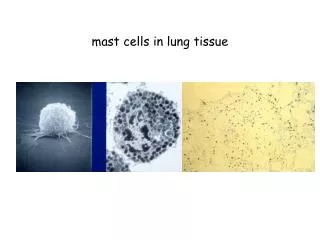
Mast cells • Derived from bone marrow precursor • Reside in tissues adjacent to blood vessels • Express a high-affinity receptor for Fc fragment of IgE • Contain numerous mediator-filled granules
Mast cells surface markers • FcεRI – high-affinity receptor for IgE • CD 117- membrane gp – mediates interaction necessary for mast cells proliferation
FcεRI • complex consists: - α- chains with two extracellular immunoglobulin domains - β- chains – four times cross the membrane – both their ends are inserted to the cytoplasm - γ- chain = homodimer - β and γ chains – contain activating motives ITAM (mediate transport of activating signal from mast cell surface after binding of IgE) in their cytoplasmic part
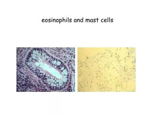
Cytoplasmic granules • Histamin (dilation of small blood vessels, increase of vascular permeability, contraction of smooth muscles), proteases (cause a damage to the tissues) • Cytokines: IL- 4 ( stimulates IgE production by B cells), 5 (activates eosinophils), 13 (stimulates mucus secretion by airway epithelial cells), TNF α, IL-1 • Growth factors: TGF- β, VEGF (vascular endothelial growth factor) • Synthetize: PAF (platelet activation factor), LTC4- leukotrienes (stimulate prolonged smooth muscle contraction), PGD2- prostaglandins (cause vascular dilation)
Mast cells function • Participate on defence against bacterial and parasites infection • Develop immediate hypersensitivity reactions (production of IgE antibodies in response to an antigen, binding of IgE to Fc receptors of mast cells, cross-linking of the bound IgE by the antigen and release of mast cell mediators) • Take part in reparation of tissues - angiogenesis, production of intercellular substances
27. Immune mechanisms of the inflammation (local and systemic reaction)
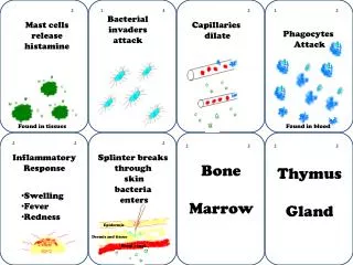
Inflammation = a complex reaction of the innate immune system in vascularized tissues that involves accumulation and activation of leukocytes and plasma proteins at a site of infection, toxin exposure or cell injury • Iniciated by changes in blood vessels that promote leukocyte recruitment and movement of fluid and plasma proteins into tissue • Inflammation serves a protective function in controlling infections and promoting tissue repair
Inflammatory response • Local - signs: redness, swelling, pain, heat • Systemic – sings: fever • Acute or chronic process
Inflammation - iniciation • 1. signal: phagocytes and degranulated mast cells, substances released from damaged cells, parts of intercellular substance exposed by injury
Local inflamation • Increase of vessels permeability – plasma fluid passes through the blood vessels- swelling • Increase of endothelial adhesivity (expression of cell adhesion molecules)- phagocytes and lymphocytes adhere to endothelial cells – pass to the tissues • The coagulation, fibrinolytic, kinin and complement systems are activated • Local nerve endings are influenced (pain) • changes in temperature regulation
Local inflamation • phagocytes- accumulate in a site of injury, produce cytokines • Ag + APC home to LN (lymph nodes) - activate T cells and induce their terminal differentiation, activation of B cells and their changes into plasma cells with production of antibodies • Mature Th 1 lymphocytes migrate from LN to the site of infection and stimulate macrophages
Systemic response (SR) to inflammation • Development of SR depends on: - range of damage - duration of local inflammation • SR can arise also without local inflammation: - after massive enter of microorganisms into the blood = septic shock - after intravascular antigen impulse of noninfection character = anaphylactic shock • Massive release of mediators (cytokines, complement, histamin)- leads to a massive vasodilatation- hypotensis- vascular collapse
Sings of systemic inflammation • Fever- caused by stimulation of hyppothalamic center of termoregulation (by proinflammatory cytokines –TNF, IL-1, INF-gama) • Caused also by increase of proteosynthesis, increase of Hsp production (hot shock protein)
Mediators of inflammation • Cytokines produced in the site of inflammation- come through the blood into the liver- stimulate production of plasma proteins of acute inflammation phase (CRP, amyloid P, complement C3,C4), transport proteins (ceruloplasmin, transferin) and inhibitors of proteases (alfa 2-macroglobulin) • Proteins of acute phase – function - opsonisation, activation of complement • Cytokines and mediators cause bone marrow to release and produce leukocytes- leukocytosis
Reparation of damaged tissues • Activation of reparation process: - occurs during later phases of inflammation - phagocytes- eliminate damaged cells • activation of fibroplastic mechanisms • activation of angiogenesis • start regeneration and remodelation of tissues • Control by cytokines, hormons, enzymes • Chronic inflammation – accompane by fibrotic process
Cytokines • Humoral factors - provide intercellular communication between immune cells + communication between immune system and other body systems • Proteins secreted by leukocytes (and by other cells of immune system) • Influence different cells of immune system through specific receptors • Forms- secreted or membrane • Effects - pleiotropic (1 cytokine has a several fysiological effects)
Function of cytokines • Activating signals for cells – activate, regulate cell cycle, mitotic activity • Cause changes of the cell membranes – increase of cytokine receptors expression • Participate in reparation of tissues in the terminal phase of inflammation • Regulate immune cells proliferation and differentiation in the immune organs • Influence migration of the immune cells
Classification of cytokines • Pro-inflammatory • Anti-inflammatory • With growth activity factor • Participating in humoral response IS • Participating in cell response IS • With antiviral effect
Proinflammatory cytokines • IL-1 – produced by macrophages and T cells; activates neutrophils, endothelial cells and T cells, induces synthesis of acute phase proteins in liver, causes fever • IL-6 – produced by T and B cells, monocytes; regulates B cell differentiation and proliferation, synthesis antibodies; stimulates hepatocytes to produce acute phase proteins • IL-8 – produced by monocytes, macrophages, endothelial cells; chemotactic factor for neutrophils • IL-12, IL-18, TNF
TNF • TNF α – produced by macrophages (+T a B cells, NK cells, neutrophiles,..) after activation by lipopolysacharids binded to plasma protein- complex binds to CD14 on the macrophage surface – causes releasing of TNF α • TNF α – participates on early phase of inflammation, induces expression of adhesive molecules on endothelial cells and leukocytes; stimulates proinflammatory protein production; stimulates catabolic processes • TNF β- produced by T and B cells; similar effects
Antiinflammatory cytokines • IL-10 – produced by Th2 cells, monocytes, macrophages, activated B cells; inhibition of cytokine Th1, Tc and NK cells production; inhibition of synthesis proinflammatory cytokines by macrophages • TGF-β – growth activator but also inhibitor of different cell types • Regulates damaged tissues reparation by stimulation of intercellular substances synthesis, modulate expression of tissue metaloproteases and tissue inhibitors of proteolytic enzymes; regulates cell adhesion; chemoatractant of fibroblasts; can inhibite T and B cell proliferation • IL-4
CYTOKINES with activity of growth factors • IL-2, IL-4, IL-5, IL-6, IL-9, IL-14 • SCF- stimulates stem cell proliferation, their release to the peripheral blood • IL-3 – influences maturation of all cell lines • IL-7 – growth factor of T cells • IL-11 – growth factor of megakaryocytes • IL-15 – induces proliferation of mast cells, Th and Tc cells
CYTOKINES with activity of growth factors • G-CSF, M-CSF, GM-CSF – stimulate granulocytes and/or monocytes/macrophages proliferation and differentiation, prolongate their survival and increase their functional capacity during inflammation • EPO = erythropoetin- stimulates red cells differentiation
CYTOKINE participating in humoral response of IS • IL-4 – produced by Th2 cells, mast cells and basophils; stimulates B cell proliferation, production of IgM,G1,E; stimulates T cell proliferation; activates macrophages; growth factor of mast cells (antiinflammatory effect) • IL-13 – see IL-4 + chemotactic factor for monocytes and macrophages • IL-5 – produced by Th2 cells, lymphocytes and mast cells; activates and stimulates B cells and eosinophils proliferation, stimulates Tc cells • IL-9 – produced by Th2 cells; co-stimulating factor - amplifies Th cells and mast cells proliferation
CYTOKINES participating in cell mediated response • IL-2 – produced by Th cells after stimulation by antigen, autocrine effect to Th cells; activating signal for Tc cells and NK cells; 2. signal for activation and differentiation of B cells • IL-12 – produced by monocytes, macrophages; stimulates maturation Th0 into Th1 cells; activates NK cells; stimulates INF- γ production by Th1 cells; inhibites IgE secretion • IFN-γ, GM-CSF
CYTOKINE with antiviral effect • Interferons- anti-inflammatory, anti-prolifferative • IFN-α, IFN-β- produced by cells with nucleus after stimulation – viruses - IFN (after binding to the receptor) induces in noninfected cell status of non-permisivity = decomposition of viral nucleus acids and influence into translation of viral proteins • IFN- α = represents 20 cytokines – differ by glycosylation of general protein structure; produced by leukocytes - in recombinant form – therapy of chronic active hepatitis B, hepatitis C
Receptors for cytokines • Consist of 2-3 subunits • 1- binds cytokine • Others mediate contact with intracellular signaling molecules • Results of signalisation: stimulation of cell mitosis, differentiation, initiation of effector functions (degranulation, secretion of cytokines, activation of membrane enzymes, chemotaxis), inhibition of cell mitosis and induction of apoptosis
Signaling process • RECEPTORS of CYTOKINES are associated with: • Protein-kinases- phosphorylate intracellular substances (after activation )- influence transcription of genes, structure of cytoskelet • G-proteins – iniciate phosphorylation (after cytokine binding to the receptor) – catalyze the replacement of GDP (guanyl diphosphate) to GTP and can activate a variety of cytoplasmic enzymes • Calcium ions – open Ca channels (after cytokine binding to the receptor) – increase of Ca ions concentration in the cytoplasma - activation of proteins by ca ions binding
Regulation of local concentration of cytokines • Biological effects are neutralized by binding to nature inhibitors and soluble forms of receptors for cytokines • MMP (matrix metaloproteases) release cytokines from molecules of intercellular substance • Elastases release binded cytokines
29. Physiological mechanisms of regulation of the immune system.
Immune mechanisms of regulation Immune system is regulated by: • Antigen • Antagonistic peptid • Antibodies • Cytokines and intercellular contact • T lymphocytes • Neuroendocrine regulation
REGULATION BY ANTIGEN • Antigen competition – peptides from different antigens compete for binding-sites on the MHC gp = antigen is able to suppress expression of other antigen IMPORTANT IS : • the binding strenght of peptide to MHC gp II • density of peptid-MHC gp II complex on the surface of APC Immune response finishes after extinction of antigen – due to a short life-span of effector lymphocytes
ANTAGONISTIC PEPTIDES • Agonists - peptide fragments of antigen with adequate binding to MHC gp, recognition by T cells with sufficient affinity – induce full response of T cells • Partial agonists – induce a qualitative different response of T cells - peptides have a similar structure, bind adequately to MHC gp, but make too weak or too strong interaction with T cells • Antagonists – induce anergy of T cells
REGULATION MEDIATED BY ANTIBODIES • Antibodies have an effector and regulatory functions • Secreted antibodies – compete with BCR for antigen = negative regulators of B cells stimulation • Immune complexes of antibody and antigen – bind to the surface of B cells – interaction of signaling molecules (protein-kinases, protein-phosphatases) = inhibit B cells activation (= antibody feedback)
REGULATION MEDIATED BY CYTOKINES AND INTERCELLULAR CONTACT • Inhibition of cytokine effect by endocytosis of their receptors or by binding of inhibitors to their receptors • Inhibitory receptors – protection against too easy T cells activation • Apoptotic receptor (Fas) mediates a negative regulation after binding to ligand FasL on activated T cells – causes – lysis of cell
NEGATIVE REGULATION mediated by T lymphocytes • Th2 cells produce IL-4, IL-10- suppress immune response of Th1 cells • Suppression of T cells occurs after recognition an antigen on cells different from APC (missing of co-stimulating signals) • CD8+ T cells – secrete soluble forms of TCR - compete with TCR on the surface of other cells- inhibition • Regulatory Tr1 cells (CD4+)- secrete IL-10-anti-inflammatory effect, induce tolerance to autoantigens
NEUROENDOCRINE REGULATION • Neurotrasmiters influence leukocytes by binding to the specific receptors (noradrenalin) • CS, growth hormons, thyroxin, endorfins – influence the leukocytes by binding to the specific receptors • Leukocytes produce endorfins, TSH, growth hormon, vitamin D3, ACTH • Cytokines influence nerve system (IL-1, IL-6) • Stress influences immune system- cortocosteroids-activity of phagocytes, NK cells
OTHER FACTORS • The same antigen can induce active immune response or active tolerance – depends on: • Condition of immune system • Character of antigen (size, structure of molecule) • Dose of antigen (too low or too high doses-induce tolerance) • Route of antigen administration (s.c.-induce immune response, p.o. or i.v.- induce tolerance)
MHC (major histocompatibility complex) • human MHC = HLA (human leukocyte antigens) • Genes of MHC complex – are codominantly expressed = alleles inherited from both parents are expressed equally; are polymorphic – many different alleles are present
Contain of MHC complex • MHC gp = are encoded by genes localized on short arm of human 6. chromosome • The MHC locus- contains 4 single blocks of loci - HLA Ags I are encoded by loci A, B, C - HLA Ags II – locus D - HLA Ags III – locus between HLA I a II (contains genes with nondirected participation in histocompatibility- genes for complement, TNF…)