Download
1 / 51
510 likes | 764 Views
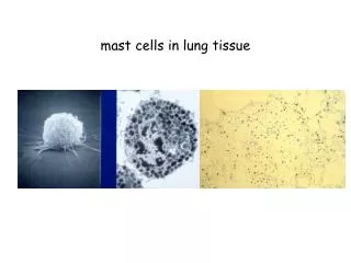
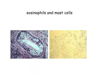
Basophils and mast cells and their importance in immune responses. Mast cells. Mucosal mast cells - in the mucous membranes of respiratory and gastrointestinal tract, produce histamine, serotonin, heparin, tryptase, leukotriene C4 ..., participate in parasitosis and allergy

E N D
Basophils and mast cells and their importance in immune responses
Mast cells • Mucosal mast cells - in the mucous membranes of respiratory and gastrointestinal tract, produce histamine, serotonin, heparin, tryptase, leukotriene C4 ..., participate in parasitosis and allergy • Connective tissue mast cells - the connective tissue, producing tryptase, chymase, prostaglandinD2 ..., are multiplicated in fibrosis, in parasitosis and allergy are not participating
Mast cell functions • Defense against parasitic infections • In pathological circumstances, responsible for the early type of hypersensitivity (immunopathological reaction typeI) • Apply during inflammation, in angiogenesis, in tissue remodeling
Mast cell activation Mast cells can be stimulated to degranulate by: • cross-linking of IgE Fc receptors • by anafylatoxins (C3a, C4a, C5a) • direct injury, alcohol, some antibiotics
Mast cell activation by cross-linking of IgE Fc receptors • Establishing of multivalent antigen (multicellular parasite) to IgE on highaffinnity Fc receptor for IgE (FcRI) • Aggregation of several molecules FcRI • Initiate mast cell degranulation (cytoplasmic granules mergers with the surface membrane and release their contents) • Activation of arachidonic acid metabolism (leukotriene C4, prostaglandin D2) • Start of production of cytokines (TNF, TGF, IL-4, 5,6 ...)
Secretory products of mast cells • Cytoplasmatic granules: hydrolytic enzymes, proteoglycans (heparin, chondroitin sulphate), biogenic amines (histamine, serotonin) Histamine causes vasodilation, increased vascular permeability, erythema, edema, itching, contraction of bronchial smooth muscle, increases intestinal peristalsis, increased mucus secretion of mucosal glands in the respiratory tract and GIT (helps eliminate the parasite) • Arachidonic acid metabolites(leukotriene C4, prostaglandin D2) • Cytokines(TNF, TGF , IL-4, 5,6 ...)
Basophils • Differentiate from myeloid precursor • They are considered to be the circulating form of mast • Receptor equipment, containing granules, the mechanisms of stimulation and functions are very similar to mast cells • They are responsible for the emergence of anaphylactic shock • basophil activation markers: CD 63, (CD 203)
Immune mechanisms of inflammation (Local and systemic reactions)
Inflammation *Is a summary of physiological responses to breach the integrity of the organism, leading to protection against infection of damaged sites, localization of damage and healing. * The first signals to the development of inflammatory responses come from mast cells, phagocytes, and the substances released from damaged cells and extracellular components of matter.
Local body's response to inflammationManifestations-pain (dolor), fever (calor), redness (rubor), swelling (tumor) and loss of function (funkcio laesa)
-increased permeability of blood vessels (vasoactive amines, complement components C3a, C5a, leukotrienes ..., swelling at site of inflammation) - increased expression of adhesion molecules on endothelia - activation of coagulation, fibrinolytic, kinin and complement system - influence of local nerve endings (prostaglandins, pain) - changes in temperature (IL-1, IL-6, TNF, prostaglandins)
Systemic response to inflammation- depends on the extent of damage and duration of local inflammation - fever (proinflammatory cytokines TNF, IL-1, IFN ; stimulate hypothalamic center of thermoregulation) - mobilization of tissue metabolism - induction of expression of Hsp (heat-shock-proteins; function as chaperones) - production of acute phase proteins (CRP, SAP, C3, C4; opsonization and complement activation) by liver after stimulation with cytokines (TNF-α, IL-1, IL-6)
-increased hepatic synthesis of certain serum transport proteins (ceruloplasmin, transferrin) - increased synthesis of protease inhibitors ( macroglobulin) - leukocytosisSeptic shock - the massive penetration of microorganisms into the bloodstream(TNF) Anaphylactic shock- basophil degranulation and complement activation with allergen (histamine)
Repair of damaged tissue - elimination of damaged cells with phagocytes - activation of fibroplastic mechanisms - activation of angiogenesis - regeneration and tissue remodeling
Regulation by antigen • Induce immune responses and extinction • Affinity maturation of B lymphocytes • Maintaining immunological memory • Antigenic competition • Threshold density of the complex MHC II-gp Ag on APC
Regulation by antagonistic peptides • Agonist - antigenic peptide, which induce full T cell response (proliferation, differentiation, TH or TC and stimulation of effector functions) • Antagonist - (partial agonist) peptide structurally similar to antigen-peptide, which induce qualitatively different response of T lymphocytes (production of only some cytokines, anergy, ...) Negative signals induced by antagonist may overcome positive signals induced by agonist (which is in the body in excess), it is used by some microorganisms
Regulation by antibodies • Antibodies competes with the BCR for antigen (negative regulator of B lymphocyte stimulating) • IgG immune complexes bind to the BCR and FcgR on B cells, resulting in blocking activation of B lymphocytes • Regulation via idiotypic network
Regulation by cytokines and cellular contact • Interaction APC - T lymphocyte • Interaction TH1 – macrophages • Interaction TH2 - B lymphocytes • Mutual regulation of activity TH1 versus TH2 • Development of leukocyte subpopulations Negative regulation of effector cells: • CTLA-4 - T cell inhibitory receptor, binds ligands CD80 and CD86 • Inhibitory receptors of NK cells • Self-destruction interaction of the apoptotic receptor Fas with ligand FasL on the surface of activated T lymphocytes
Interaction of APC with T lymphocyte T cell:TCR - antigen-specific receptor (signal 1)CD4 or CD8 - coreceptor (MHCgp binding)CD 28 - costimulatory receptor (signal 2, binds CD 80, CD 86)CTLA-4 - inhibitory receptor (binds CD 80, CD 86)CD-40LAPC:MHC gp I + antigenic peptideMHC gp II + antigenic peptideCD 80, CD 86 - costimulatory ligandsCD 40
Suppression mediated by T lymphocytes • Mutual negative interaction TH1 and TH2 cytokine-mediated (TH2 lymphocytes produce IL-4 and IL-10 that suppress the immune response based on TH1 cells) • CD 8+ TS - suppressor T cells has not yet been isolated as a separate subset (partly identical with TC) - negatively regulate the activation of other T cells • Soluble suppressor factors - some CD 8+ T lymphocytes produce a soluble form of TCR • Clonal elimination or anergy of T lymphocytes after contact with antigen on the surface of other cells than APC (lacking costimulating signals) • Regulatory T cells (Tr1 CD 4+) help to maintain tolerance to autoantigens
Factors influencing the outcome of the immune response The same antigen can induce an active immune response or an active state of tolerance, the result of response depends on many factors: • State of the immune system • Properties of antigen • Dose of antigen • Route of antigen administration
Cytokines • Regulatory proteins and glycoproteins produced by leukocytes and other cells • Essential regulators of the immune system • Apply also outside the immune system (angiogenesis, tissue regeneration, carcinogenesis, treatment of many brain functions, embryonic development ...) • Cytokines - secreted - membrane (CD 80, CD86, CD40L, FasL ..)
Pleiotropic effect • Operates in a cascade • Cytokine Network • Cytokine system is redundant • Effects of cytokines - autocrine - paracrine - endocrine • Are known as interleukins (exception: TNF, lymphotoxin, TGF, interferons, CSF and growth factors)
B cells communicate via cytokines with other inflammatory cells, such as T cells and macrophages
Distribution of cytokines by function • Proinflammatory cytokines (IL-1, IL-6,IL- 8,IL- 12,IL- 18, TNF) • Antiinflammatory cytokines (IL-1Ra, IL-4, IL-10, TGF) • Cytokines with the activity of hematopoietic cells growth factor (IL-2, 3, 4, 5, 6, 7, 9, 11, 14, 15, CSF, SCF, LIF, EPO) • Cytokines applying in TH2 humoral immunity (IL-4, 5, 9, 13) • Cytokines applying in the cell-mediated immunity TH1 (IL-2, 12, IFN, GM-CSF, lymphotoxin) • Cytokines with anti-virus effect (IFN-, IFN-, IFN- )
Cytokine receptors • Consisting of 2 or 3 subunits • One subunit binds cytokine, other are associated with cytoplasmic signaling molecules (protein kinases) • Signaling subunit is shared by several different cytokine receptors - called receptor family • Signaling through these receptors may lead to proliferation, differentiation, activation of effector mechanisms or blocking the cell cycle and induction of apoptosis
MHC glycoproteins class I (Major histocompatibility complex) • The function of MHCgpI is presentation of peptide fragments from inside the cell (which are produced by cell, including viral peptides if are present) on the cell surface so to T lymphocytes (cytotoxic CD8+) • Present on all nuclear cells of the organism • 3 isotype classical human MHC gp.(HLA - A,-B,-C) • 3 isotype-classical MHC gp.(HLA - E,-F,-G; molecule CD1)
Structure of MHC gp I • MHC gp class I consists of transmembrane chain a and non-covalently associated B2mikroglobulin • a chain has 3 domains, 2 N-terminal (a1, a2 - binding site for peptides) and 1 C-terminal domain (a3 - anchored in the cytoplasmic membrane, a structure similar to imunoglobulin domain) • Binding of peptide is necessary for a stable conformation of MHCgp and thus ensure its long presentation on the cell surface
Peptides binding to MHCgpI • MHC gp I bind peptides with a length of 8 to 10 aminoacides • Certain MHC gp molecule binds peptides sharing common structural features - coupling motif (critical are aminoacides near the end of peptide) • The binding of endogenous peptides occurs in the endoplasmic reticulum during biosynthesis of MHC gp • After a string a and b2mikroglobulin create in the ER, folding into the correct conformation and the mutual association and the association of an appropriate peptide, the complex is further processed in the Golgi apparatus and then is presented on the cell surface • Linked peptides derived from proteins degraded proteasome, which cleaves cytoplasmic proteins for destruction (labeled with ubiquitin), peptide fragments are transported into the ER by specific membrane pump
Non-classical MHC gpI • HLA - E,-F,-G; CD1 molecules • Structurally similar to classical MHC gp • Are less polymorphic • There are only on some cells • They specialize in binding of specific ligands
HLA-E and HLA-G - occurs on the trophoblast cells • Complexes of HLA-E and HLA-G with peptides are recognized by inhibiting receptors of NK cells and contribute to the tolerance of the fetus in utero • CD1 molecules - bind glycolipids (recognized by NK-T lymphocytes)
MHC glycoproteins class II • The function of MHC gpII is the presentation of peptide fragments from protein whitch were engulfed by antigen presenting cell on the cell surface to T lymphocytes (auxiliary CD4) • Occur on the APC (dendritic cells, monocytes, macrophages, B lymphocytes) • 3 isotypes of MHC gpII (DR, DQ, DP)
Structure of MHC gp II • MHC gp II consist of 2 non-covalently associated transmembrane subunits a and b • The peptide binding site consists of N-terminal domains a1 and b1 • Binding of peptide is necessary for a stable MHC gp conformation and thus ensure its long presentation on the cell surface
Binding of peptides to MHC gp II • MHC gpII bind peptides with a length of 15 to 35 aminoacides (but possibly longer - because the peptide binding site is open at both ends) • Certain MHC gp molecule binds peptides sharing common structural features - coupling motif • After a string a and b are created in ER, fold into the correct conformation and the mutual associated are connected with another transmembrane chain called invariant chain, which blocks the binding site for the peptide, this complex is further processed in the Golgi apparatus, secretory vesicles isolated from GA merge with endosomes, then split the invariant chain and peptide fragments from cell absorbed proteins bind into binding site of MHC gp and the complex is then presented on cell surface
Polymorphism of MHC glycoproteins • HLA complex is located on chromosome 6 • For MHC gp is typical high polymorphism (except the non-classical MHC gp, and DR a chain) • Codominant inheritance of alelic forms (Individual has 3 cell surface isotypes of HLA molecules (HLA-A,-B,-C) mostly in 2 different alelic forms) • Polymorphism has a protective significance at individual and population level • Ppolymorphism MHC gp causes complications in transplantation
HLA typing = determmination of HLA antigens on the surface of lymphocytesCarry out during the testing before transplantation and in determination of paternity 1) Serotyping - Microlymfocytotoxic test Allospecific serums (obtained from multiple natal to 6 weeks after birth, obtained by vaccination of volunteers, or commercially prepared sets of typing serums (monoclonal antibodies)) Principle - the incubation of lymphocytes with typing serums in the presence of rabbit complement, then is added the vital dye which stained dead cells - cells carrying a specific HLA are killed by cytotoxic Ab against the Ag, the percentage of dead cells is a measure of serum toxicity (forces and antileukocyte antibody titre) Positive reaction is considered more than 10% dead cells (serological typing can be done also by flow cytometry
2) Molecular genetic methods • For typing are used hypervariable sections in the II. exon genes coding for HLA class II; to determine HLA class I is used polymorphism in II. and III. exon coding genes2a) PCR-SSP= Polymerase chain reaction with sequential specific primers • Extracted DNA is used as a substrate in a set of PCR reactions • Each PCR reaction contains primers pair specific for a certain allele (or group of alleles) • Positive and negative reactions are evaluated by electrophoresis, each combination of alleles has a specific electrophoretic painting
2b) PCR-SSO PCR reaction with sequence-specific oligonucleotides Multiplication of hypervariable sections of genes coding HLA Hybridization with enzyme or radiolabeled DNA probes specific for individual alleles 2c) PCR-SBT Sequencing based typing The most accurate method of HLA typing. We get the exact sequence of nucleotides, which compares with a database of known sequences of HLA alleles