Download
1 / 72
730 likes | 751 Views
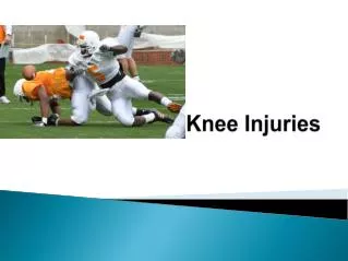
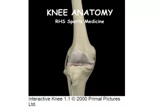
Anatomy and Injuries of the Knee. Adapted from Connie Rauser Sabino Sports Medicine. Bones Femur Medial/lateral femoral condyles articulate w/ tibia Tibia Tibial plateau is flat-articulates w/ femoral condyles Fibula Articulates w/ tibia Patella Sesamoid bone protects anterior joint

E N D
Anatomy and Injuries of the Knee Adapted from Connie Rauser Sabino Sports Medicine
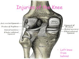
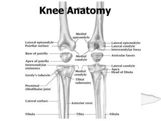
Bones • Femur • Medial/lateral femoral condyles articulate w/ tibia • Tibia • Tibial plateau is flat-articulates w/ femoral condyles • Fibula • Articulates w/ tibia • Patella • Sesamoid bone protects anterior joint • Enclosed in quadriceps/patellar tendon Anatomy-Bones
Joints • Tibiofemoral • Hinge joint with synovial lining • diarthrodial • Patellofemoral • Superior Tibiofibular Anatomy-Joints
Meniscus • Medial and lateral • Fibrocartilaginous disks • Thicker on outside than inside (poor blood supply) • Lie on top of tibial plateau • Increase stability • Make condyles fit better • Shock absorbers Anatomy-Meniscus
ACL-anterior cruciate ligament • Runs from anterior tibia to posterior femur • Prevents anterior displacement of tibia on fixed femur • Prevents femur from moving posterior during weight bearing • Stabilizes tibia against excessive internal rotation Anatomy-Ligaments
PCL-posterior cruciate ligament • Runs from posterior tibia to anterior femur • Prevents posterior translation of tibia on fixed femur • Prevents femur from moving anterior during weight bearing • Both ACL and PCL “cross” or wrap around each other—taut when in extension and looser when in flexion Ligaments
MCL-medial collateral ligament • Attaches on the medial femoral epicondyle & anteromedial tibia • Thickened portion of joint capsule • Two parts-superficial and deep • Deep portion attaches to medial meniscus • Stabilizes against valgus stress applied to lateral aspect of joint capsule Ligaments
LCL-lateral collateral ligament • Attaches to lateral femoral epicondyle and head of fibula • Stabilizes against varus stress when force is applied to medial aspect of joint • Both the MCL and LCL are tightest during full extension of knee and relaxed during flexion Ligaments
Quadriceps • Rectus femoris, vastus lateralis, vastus medialis, vastus intermedius • Knee extension, hip flexion • Hamstrings • Biceps femoris, semimembranosus, semitendinosus • Knee flexion, hip extension Muscles
Gracilis • Knee flexion, hip adduction • Sartorius • Knee flexion, hip flexion, hip external rotation • Popliteus • Knee flexion • Gastrocnemius • Knee flexion Muscles
Plantaris • Knee flexion • Pes anserine • Goose’s foot • Knee flexion, some internal rotation • Gracilis, sartorius, semitendinosus • Iliotibial Band • Thick band on lateral aspect of thigh • Attaches at Gerdy’s tubercle on the lateral aspect of tibia Muscles
Conditioning • Strength, flexibility, cardiovascular and muscular endurance • Hamstring strength 60% of quad strength • Rehabilitation • Strengthen all muscles around knee joint • Shoes • proper type for surface • Length of cleats • Turf vs grass Preventing knee injuries
Knee braces • Functional vs. prophylactic • Functional—used to provide support to an unstable knee • Usually custom fitted to some degree • Uses hinges and supports to control excessive rotational stress and tibial translation • Prophylactic-worn on lateral aspect knee to protect MCL. • Usefulness questioned—does it cause more injuries? Preventing knee injuries
MOI: • fixed foot and external rotation of femur • knee in valgus position • hyperextension • S/S: • “pop”, • knee gives out • instability of knee joint • swelling within knee joint—hemarthrosis • intense pain initially but still able to walk • “+” Lachman’s test • “+” anterior drawer test ACL rupture
Hyperextension MOI
The ACL intact The ACL torn Inside the knee joint
Tx: RICE, knee immobilizer, crutches, Physician referral • Requires surgical reconstruction • Timing of surgery decided by athlete, parents, doctor • Grafts used are patellar tendon, hamstring tendon, cadaver graft, allograft • 3-5 weeks in brace, 6-9 months return to activity ACL Rupture
Knee post-ACL tear • Test for Swelling • Ballotable Patella Test ACL Rupture
Lachman’s test Stress tests
Modified Lachman’s Stress tests
Anterior Drawer test Stress tests
MOI: • hyperflexion • falling on bent knee with foot plantar flexed • Hit on fixed anterior tibia • S/S: • “pop” at the back of knee • Pt. Tender and swelling in popliteal fossa • + posterior sag test,+ posterior drawer test PCL Rupture
Tx: • RICE • Immobilization • Crutches • Physician referral • 6-8 weeks rest/rehab • If surgery is elected, 6 weeks immobilization PCL rupture
Posterior sag Stress tests
Sunrise or posterior sag Stress tests
MOI: • Blow to the lateral side of knee (valgus stress) • External rotation of tibia MCL Sprain
2nd degree?? MCL sprain
S/S: • 1st degree • Pt. Tender over MCL, stable but pain with valgus stress, mild joint effusion, mild joint stiffness, full ROM • 2nd degree • Partial tearing-superficial portion, Pt. Tender over MCL, some instability with valgus stress but solid endpoint, moderate joint effusion, joint stiffness, limited ROM, unable to fully extend knee joint MCL sprain
S/S: • 3rd degree • Complete tear—superficial and deep portions • Pt. Tender over MCL • Moderate to severe effusion • Severe pain • Loss of motion due to pain, effusion, muscle guarding • “+” valgus stress in 0 and 30 degrees, no endpoint MCL Sprain
Valgus stress test @ 0 Valgus stress @ 30 Stress tests for MCL
Tx: • RICE • Crutches • Knee immobilizer/brace • 1st degree 1-2 weeks • 2nd degree 2-4 weeks • 3rd degree 4-6 weeks • Physician referral for 2nd degree or greater MCL Sprain
The terrible triad or unhappy triad • Torn ACL • Torn MCL • Torn Medial meniscus Complications
MOI: • Varus force to medial aspect of knee • internal rotation of tibia • S/S: • Pt. Tender over LCL, • pain, • swelling, • loss of motion, • “+” varus stress at 30 degrees—solid endpoint with 1st degree, less stability but solid endpoint with 2nd degree, no endpoint with 3rd degree • if “+” varus stress at 0 degrees flexion suspect ACL or PCL injury as well LCL sprain
Tx: • RICE • Crutches • Knee immobilizer • Physician referral with 2nd or 3rd degree LCL sprain
Medial: more often torn than later due to attachment to MCL • Lateral: doesn’t attach to joint capsule making it more mobile, less prone to injury • MOI: • Weight bearing with rotational force while extending or flexing the knee Meniscus tear
S/S: • Effusion w/in 48-72 hours • Pt. Tender over joint line • Loss of motion • “locking” • Giving out • Pain with deep knee flexion--squatting Meniscus tear
Types of meniscus tears Meniscus tear
McMurray Test • Positive Sign: Pain and/or clicking Meniscus Tears Special Test
Tx: • RICE • Crutches if necessary • Physician referral • If knee is “locked” by displaced meniscus, go to ER • Arthroscopic surgery to fix Meniscus tears
Dislocation • Subluxation • Fracture • Chondromalacia • Patellar tendonitis Injuries to the Patella
MOI: • Foot planted, deceleration, and cutting in opposite direction from the weight bearing foot • Thigh rotates internally while leg rotates externally • Strong forceful contraction of quads (vastus lateralis) Patella Dislocation
S/S: loss of motion/function at the knee • Pain • Swelling • Deformity • Pt. Tender over medial aspect of knee joint Dislocation
Tx: • immobilize in position you find it • Ice • ER visit • After reduction, immobilize in extension about 4 weeks—use crutches • Strengthen muscles of knee, thigh and hip Dislocation