Download
1 / 37
590 likes | 792 Views
KNEE ANATOMY. RHS Sports Medicine. BONY ANATOMY OF THE KNEE. TIBIA. FIBULA. 3. 4. FEMUR. PATELLA. 1. 2. PALPATE THE LATERAL JOINT LINE. PALPATE THE MEDIAL JOINT LINE. PALPATE THE HEAD OF THE FIBULA. KNEE JOINT LINE.

E N D
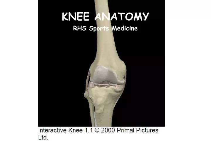
KNEE ANATOMY RHS Sports Medicine
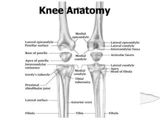
BONY ANATOMY OF THE KNEE TIBIA FIBULA 3 4 FEMUR PATELLA 1 2
PALPATE THE LATERAL JOINT LINE. • PALPATE THE MEDIAL JOINT LINE • PALPATE THE HEAD OF THE FIBULA KNEE JOINT LINE • The joint line of the knee is where the tibia and the femur meet.
TIBIA • The tibia is main weight bearing bone of the lower leg. • Theproximalend of the tibia is part of the knee joint. • The top of the tibia is flat and is called the tibial plateau. • LABEL TIBIA ON YOUR WORKSHEET. • CIRCLE THE TIBIAL PLATEAU ON YOUR WORKSHEET.
FIBULA • The fibula is a NON- weight bearing bone on the lateral aspect of the lower leg. • Theproximal end of the fibula is near the knee joint but is not considered an actual part of the joint. • LABEL FIBULA ON YOUR WORKSHEET.
FEMUR • A common name for the femur is the “thigh bone”. • The femur is the longest, largest, strongest bone in your body. • When you stand or walk, all the weight of your upper body rests on them. • The femur is part of the knee joint distally and the hip joint proximally. LABEL THE FEMUR ON YOUR WORKSHEET
PATELLA • The common name for the patella is the “kneecap”. • The patella is an irregularly shaped sesamoid bone. • The anterior surface is smooth and rounded. • The posterior surface is wedge-shaped. • It is possible to break your kneecap!
FEMUR FEMUR PATELLA PATELLA TIBIA TIBIA FIBULA FIBULA BONES OF THE KNEE JOINT ? ? ? ?
KNEE MOTIONS EXTENSION: Straightening the leg Quadriceps Muscles FLEXION Bending the knee Hamstring Muscles
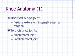
KNEE MOTION • The knee joint is a hinge joint. • Flexion and extension happens between the femur and the tibial plateau. • The patella articulates with the femoral groove.
PATELLAR TENDON The patellar tendon attaches the patella to the tibial tuberosity. DISTALLY, all Quadriceps muscles attach firmly to the superior edge of the patella.
(ANTERIOR VIEW) KNEE LIGAMENTS • Ligaments run from bone to bone. There are 4 main ligaments in the knee. • Ligaments HOLD bones together. • Ligaments are NOT involved in moving a joint! • They have different shapes. Some ligaments are like cords (lateral). Some are thin and wide (medial). (POSTERIOR VIEW)
KNEE LIGAMENTS – MEDIAL (MEDIAL VIEW) Medial Collateral Ligament (MCL) • Attaches to the femur and to the tibia. • 2 layers Thick and flat Outer = longer Inner = shorter Stronger than lateral ligament and more likely to be injured
(LATERAL VIEW) (ANTERIOR VIEW) KNEE LIGAMENTS - LATERAL Lateral Collateral Ligament (LCL) • Attaches to the femur and the head of the fibula. • Easily palpated along the lateral aspect of the knee when knee is extended. • Cord-like in shape. Less likely to be injured
LCL MCL KNEE LIGAMENTS: INTERNAL ACL A nterior C ruciate L igament femur The ACL attaches to the femur and to the tibia in the center of the knee joint. It attaches posteriorly on the femur but angles forward to attach at the front of the tibial plateau. tibia
KNEE LIGAMENTS: INTERNAL ACL Keeps the tibia from moving forward on the femur (Anterior Translation)
KNEE LIGAMENTS: INTERNAL PCL P osterior C ruciate L igament femur The PCL attaches in the opposite direction of the ACL - anteriorly on the femur but angles backward to attach over the edge and in the back of the tibia. It crosses the ACL inside the knee joint. tibia
KNEE MUSCLES QUADRICEPS (4) Rectus femoris Underneath(Vastus Intermedius) Vastus Medialis Vastus Lateralis Major Motion: Extension of the Knee
QUADRICEPS #1 Rectus femoris • Muscle starts on Pelvis • Ends at top of Patella • Crosses two joints
QUADRICEPS #2 Vastus intermedius Underneath the rectus femoris • Muscle starts along the lateral edge of the femur. • Ends at top of Patella. • Hidden underneath the rectus femoris. Front View
QUADRICEPS #3 Vastus Medialis • Muscle starts along the medial edge of the femur. • Ends at top of Patella. • Upper part hidden under other muscles. • Can be palpated near the knee. Front View
QUADRICEPS #4 Vastus Lateralis • Muscle starts along the lateral edge of the femur • Ends at top of Patella. • Upper part hidden under other muscles. • Can be palpated near the knee. Front View
QUIZ - QUADS Rectus femoris (Vastus Intermedius) Vastus Lateralis Vastus Medialis Major Motion: Extension of the Knee
KNEE MUSCLES HAMSTRINGS (3) Biceps femoris Semitendonosus Semimembranosus Major Motion: Flexion of the Knee
HAMSTRING #1 Biceps femoris • Muscle has two starting points: • Ishial Tuberosity (bone you sit on) and along back of femur • Ends at Head of Fibula • Crosses two joints
HAMSTRING #2 Semitendonosus • Muscle originates on the Ishial Tuberosity • Ends at medial edge of tibia. • Inserts over the top of the semimembranosus, almost a common tendon.
HAMSTRING #3 Semimembranosus • Muscle originates on the Ishial Tuberosity • Ends at medial edge of tibia. • Inserts close to semitendonsus, almost a common tendon.
ARTICULAR CARTILAGE • Articular cartilage lines the ends of the bones (tibia, femur) and the undersurface of the patella. • Articular cartilage protects the ends of the bones from the forces of running, jumping and walking.
ARTICULAR CARTILAGE • Articular cartilage can be torn or crushed by a force injury, such as a sudden hard step or a twisting motion where the femur twists on top of the tibia. • Articular cartilage can also be worn down by use over time, exposing the bone to the forces of motion.
MENISCUS • Two half-moon shaped pieces of cartilage that lie between surfaces of the femur and the tibia. • The meniscus (menisci) absorb the large amount of shock that occurs when the bones crash together while walking, running and jumping.
MENISCUS • The menisci also provide a “cradle” for the femur to sit atop the tibia instead of slipping off when moving. • This “cradle” helps to keep the knee joint stable,
MENISCUS • The meniscus is for the most part “avascular”, meaning it doesn't have blood vessels throughout the tissue. • Only the outermost 20% of the meniscus has a blood supply. As a result, a torn meniscus doesn't have the ability to heal itself unless there is just a small tear confined to the peripheral vascular zone.
Thicker outside edge • More “C” shaped • Bigger diameter • Thinner MENISCUS • MEDIAL MENISCUS • LATERAL MENISCUS