Download
1 / 40
410 likes | 737 Views
Female reproductive syste m. Female Reproductive Anatomy. Ovaries are the primary female reproductive organs Make female gametes (ova) Secrete female sex hormones (estrogen and progesterone) Accessory ducts include uterine tubes, uterus, and vagina

E N D
Female Reproductive Anatomy • Ovaries are the primary female reproductive organs • Make female gametes (ova) • Secrete female sex hormones (estrogen and progesterone) • Accessory ducts include uterine tubes, uterus, and vagina • External genitalia – external sex organs
Ovaries • Ovaries contain the ovarian follicles • Each follicle consists of an immature egg (oocyte) • Cells around the oocyte are called: • Follicle cells (one cell layer thick) • Granulosa and theca cells (when more than one layer is present) • The follicles and the oocytes are going through cyclic development – ovarian cycle
The ovarian cycle • Combination of follicles and oocyte development • Divided to 2 major periods (phases) • Follicular phase – period of follicle growth (days 1–14) • Luteal phase – period of corpus luteum activity (days 14–28) • The 2 phases are “separated” by Ovulation (release of the secondary oocyte from a tertiary follicle)
Luteal phase Follicular phase ovulation http://biology.clc.uc.edu/courses/bio105/sexual.htm
Follicular development - Folliculogenesis • The folliculogenesis occurs during follicular phase • Primordial Follicle –flattened granulosa cell layer, basement membrane, oocyte • Primary Follicle – growth of oocyte, zonapellucida formation, cuboidal granulosa cells • Secondary Follicle – add layers of granulosa cells, formation of theca cells
Folliculogenesis • Early Tertiary Follicle – antrum formation, zona pellucida thickens, theca interna and theca externa form, basement membrane is still present between theca and granulosa cells, blood vessels are in the theca cell layer but not in follicle • Tertiary/pre-ovulatory/Graffian – full size follicle ready to ovulate; oocyte surrounded by corona radiata (granulosa cells) and attached to follicular wall by the comulus oophorus
Follicular Phase • A few follicles begin to develop from primordial follicle • Oocyte grows, granulosa cells proliferate • Zona pellucida and antrum form
Follicular Phase • Dominant follicle continues development, rest regress • Corona radiata develops • Graafian follicle = mature follicle • Ovulation
Luteal Phase • After ovulation, the ruptured follicle collapses, granulosa cells enlarge, and along with internal thecal cells, form the corpus luteum • The corpus luteum secretes progesterone and estrogen • If pregnancy does not occur, the corpus luteum degenerates in 10 days, leaving a scar (corpus albicans) • If pregnancy does occur, the corpus luteum produces hormones until the placenta takes over that role (at about 3 months)
Luteal Phase • Ruptured follicle gland = corpus luteum • Corpus luteum secretes mostly progesterone • Corpus luteum reaches max activity 10 days, then degenerates
Corpus luteum • The fate of the corpus luteum depends on fertilization: • If pregnancy does not occur, the corpus luteum degenerates in 10 days, leaving a scar (corpus albicans) • If pregnancy occurs, the corpus luteum produces hormones until the placenta takes over that role (at about 3 months)
Oogenesis – oocyte development FEMALE STAGE OF CELL DIVISION MALE Spermatogonium 1 MITOSIS Oögonium Germ cell proliferation 46 chromosomesper cell (only twoshown here) Embryo Embryo 46(diploid) Oögonia Spermatogonia MEIOSIS 2 DNA replicatesbut no cell division. Primaryoocyte Primaryspermatocyte Sisterchromatids Sisterchromatids 46 chromosomes,duplicated 3 First meioticdivision First polarbody Secondaryoocyte(egg) Reproductive adult Secondaryspermatocytes Primary gamete dividesinto two secondary gametes. 23 chromosomes,duplicated (may notoccur) Reproductive adult 4 Second meioticdivision Spermatids Disintegrates Egg releasedfrom ovary atovulation. Secondary gamete divides. develop into 23 chromosomes(haploid) Sperm FERTILIZATION 6 One primary oocyteyields 1 egg. One primary spermatocyteyields 4 sperm. 5 Secondpolar bodydisintegrates. Unfertilized eggpasses out of body. Zygote
Oogenesis • Ovum production • Occurs monthly in ovarian follicles • Part of ovarian cycle • Happens during the Follicular phase (preovulatory)
Oogenesis • Production of female sex cells by meiosis • In the fetal period, oogonia (2n ovarian stem cells) multiply by mitosis and store nutrients • Primordial follicles appear as oogonia are transformed into primary oocytes • Primary oocytes begin meiosis but stall in prophase I
Oogenesis: Puberty • At puberty, one activated primary oocyte produces two haploid cells • The first polar body • The secondary oocyte • The secondary oocyte arrests in metaphase II and is ovulated • If fertilized, the second oocyte completes meiosis II, yielding: • One large ovum (the functional gamete) • A tiny second polar body
Oogonia(multiple by mitosis until 5th month of fetal development) Arrested development (until shortly before birth) Primary oocytes (arrest in prophase I) ___________________________________________________________ Puberty Oocyte in Graafian follicle – complete meiosis I Secondary oocyte first polar body Arrested in metaphase II If fertilization occur Complete meiosis II Ovum second polar body
Uterus • Hollow, thick-walled organ located in the pelvis anterior to the rectum and posterosuperior to the bladder • Body – major portion of the uterus • Fundus – rounded region superior to the entrance of the uterine tubes • Cervix – narrow neck which projects into the vagina inferiorly • Cervical canal – cavity of the cervix that communicates with: • The vagina via the external os • The uterine body via the internal os
Uterine Wall • Composed of three layers • Perimetrium – outermost serous layer; the visceral peritoneum • Myometrium – middle layer; smooth muscle • Endometrium – mucosal lining of the uterine cavity
Endometrium • Has numerous uterine glands that change in length as the endometrial thickness changes • Stratum functionalis: • Undergoes cyclic changes in response to ovarian hormones • Is shed during menstruation • Stratum basalis: • Forms a new functionalis after menstruation ends • Does not respond to ovarian hormones
Uterine Tubes (Fallopian Tubes) and Oviducts • Receive the ovulated oocyte and provide a site for fertilization • Empty into the uterus via the isthmus • Expand distally around the ovary forming the ampulla • The ampulla ends in the funnel-shaped, ciliated infundibulum containing fingerlike projections called fimbriae • The uterine tubes have no contact with the ovaries • Beating cilia on the fimbriae create currents to carry the oocyte into the uterine tube • The oocyte is carried toward the uterus by peristalsis and ciliary action
The Uterine tubes • Uterine tubes (Fallopian tubes or oviducts) • Infundibulum • End closest to the ovary with numerous fimbriae • Ampulla • The middle portion, widest and longest portion • Isthmus • A short segment connected to the uterine wall • The mucosa of the uterine tube has ciliated simple columnar epithelial cellsthat beat toward the uterus and, with the help of muscular contractions of the tube, transmit the egg in that direction. • Fertilization occurs in uterine tube (ampulla)
Uterine cycle • Repeating series of changes in the endometrium • Menses • Degeneration of the endometrium • Menstruation • Proliferative phase • Restoration of the endometrium • Secretory phase • Endometrial glands enlarge and accelerate their rates of secretion
Beginning of new cycle - Menstrual Phase of the Uterus • If fertilization does not occur, the corpus luteum degenerates and estrogen and progesterone levels decrease. • The lack of estrogen and progesterone leads to the collapse of the endometrium, which in turn leads to menstruation.
Menstrual and prolifarative phases (corresponds Follicular Phase of Ovary) • FSH and LH increase during follicular phase because progesterone concentration is low and therefore negative feedback on these pituitary hormones is low. • FSH and LH stimulate primary follicles (containing primary oocytes) to grow and stimulate their theca cells to produce estrogen. • Estrogen leads to a thickening of the endometrium.
Menstrual and prolifarative phases (corresponds Follicular Phase of Ovary) • The one dominant follicle (Graafian follicle) survives because • it is hyperresponsive to FSH and can maintain itself even under low FSH • it also becomes sensitive to LH. • LH surge appears because increased estrogen exerts a positive feedback effect on the LH releasing mechanism of pituitary. • LH surge leads to release of the primary oocyte (ovulation)
Secretory Phase of the Uterus (corresponding to Luteal Phase of Ovary ) • The now empty follicle, corpus luteum, starts secreting progesterone that exert a negative feedback on secretion from LH and FSH, preventing new follicles from maturing. • Progesterone converts the endometrium into a secretory tissue full of glycogen and blood vessels, ready to receive a fertilized egg.
The Uterine Cycle Figure 26-13 (2 of 2)
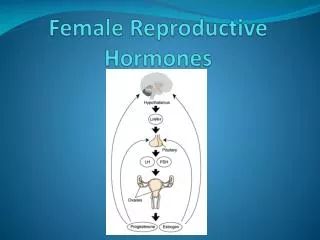
Complex Hormonal Control of Two Cycles • Ovarian and uterine cycles are controlled by several hormones, which display complex interactions • Extra-ovarian hormones • GnRH from the hypothalamus • FSH and LH from the anterior pituitary • Ovarian hormones • Estrogen • Progesterone • Inhibin
Corpus Luteum Degenerates and Ceases Hormone Production Phases of theOvarian Cycle Luteal Phase Ovulation Follicular Phase LH Gonadotrophichormonelevels FSH Ovariancycle Corpusluteumformation Maturecorpusluteum Primaryfollicle Corpusalbicans Ovulation Theca Antrum Progesterone Ovarianhormonelevels Estrogen Inhibin Uterinecycle PROLIFERATIVEPHASE Phases of theUterine Cycle MENSES SECRETORY PHASE 36.7 Basal bodytemperature (–C) 36.4 Figure 26-13 (4 of 4) DAYS 28/0 7 14 21 28/0
Hormonal Control of the Menstrual Cycle: Late Luteal Phase LH FSH Ovum Follicle Corpus luteum Estrogen Inhibin Progesterone GnRH GnRH GnRH GnRH Hypothalamus Pituitary Tonic secretionresumes FSH LH FSH LH FSH LH FSH LH Corpus luteum(from ovulatedfollicle) Corpusluteumdies Follicle New folliclesbegin todevelop Follicle Thecalcells Granulosacells secretes Granulosacells Thecalcells Estrogen Androgens Inhibin Estrogen andprogesterone Progesterone Androgens Inhibin High estrogenoutput Small amount ofprogesterone Estrogens (a) Early to mid-follicular phase (b) Late follicular phase and ovulation (c) Early to mid-luteal phase (d) Late luteal phase Figure 26-14 (4 of 4)
Mammary Glands • Pectoral fat pad • Modified sweat glands consisting of 15-25 lobes that radiate around and open at the nipple • Areola – pigmented skin surrounding the nipple • Lobes contain glandular alveoli that produce milk in lactating women • alveolar glands pass milk to lactiferous ducts, which open to the outside
Mammary Glands • nonlactating breast consists mostly of adipose and collagenous tissue and contains very little mammary gland. • Lactating • develops during pregnancy, • it exhibits 15–20 lobes arranged around the nipple, each drained by a lactiferous duct. These dilate to form a lactiferous sinus opening into the nipple.